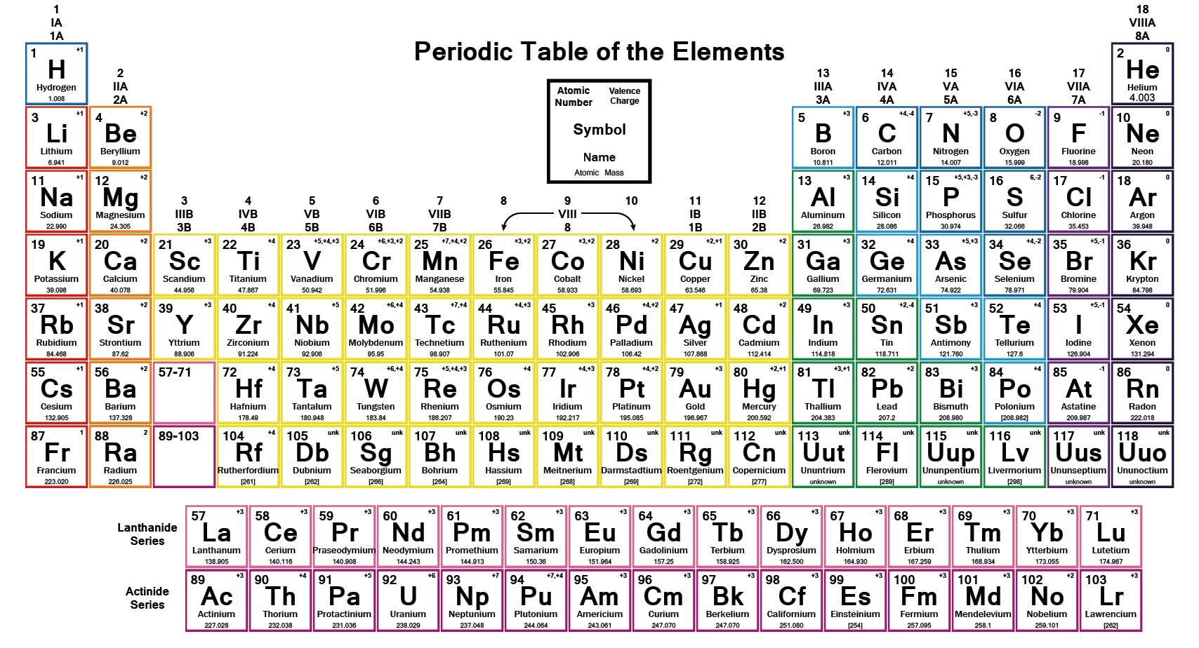
What are the essential metals for human health? Are these metals also found in plants we eat? How are they transported from the gut, or soil, into our blood, or plant? Let’s start with a review by Maria Antonietta Zoroddu and her fellow scientists from Italy and Norway. These scientists identified nine. Selenium is not technically a metal, but we’ll add it to their list. A column has been added to the table if it is in this Centrum multivitamin.
Metal
Centrum
Charge
curcumin
tannic acid
quercetin family
p-coumaric acid
e- transfer
Na,
+1
+1
no
K
2%
+1
+1
no
Mg
24%
+2
+2
+2
+2
no
Ca
16%
+2
+2
+2
no
Fe
33%
+2, +3
+2, +3
+2, +3
+2, +3
yes [2]
Mn
100%
+2, +4, +7
+2
+2
+2
yes [2]
Co
+2, +3
+2
+2
+2
+2
yes [2]
Cu
100%
+1, +2
+2
+2
+2
+2
yes [2]
Zn
100%
+2
+2
+2
+2
no
Mo
111%
+4, +6
+2
+6
yes [2]
Se
182%
-2, +4
yes [3]
This table combines essential metals for human health [1], comments on thier ability to transfer electrons [2,3], and four exemplary plant polypenols that chelate these metals from ref [4]
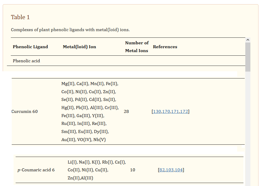
A review by Fedenko and coauthors provides an excellent analysis of plant polyphenolics that can bind metals and metalloids. [4] This table provides some of the phenolics that readers might be familiar and the oxidation state of the metal, metalloid bound.
These are some screen shots from the Fedenko 2022 review [4] Figure 1. The reader of this post is invited to consult this public access review to toxic metal binding properties of plant polyphenols. The Fedenko review contains a massive amount of peer reviewed data. It is interesting to note that no review mentioned Cu(I) binding. It is not know if this is because Cu(I) was not tested or if it was not considered toxic in the first place. Certainly Fe(II) and Fe(III) was tested in many of the polyphenols referenced by Fedenko and coauthors. [4]
These are some structures of polyphenols from PubChem mentioned in this post. Oxygens are red and carry a partial, if not an outright, negative charge for binding cations. Judging from table 1 of the Fedenko review, the tendency is to bind di- and tri- valent cations.
The philosophical twist
Metal ions may perform structural as well as catalytic roles in proteins of our bodies. [3] Often the catalytic roles involve the well coordinated transfer of electrons from one place to another. Metals may become toxic if they transfer that electron to O2 to form reactive oxygen species. Plant polyphenols have a way of binding metals that are used for legitimate purposes in enzymes as well as environmental contaminates such as cadmium Cd(II) and lead Pb(II). Plants are being considered agents of bioremediation to clean up environmental contaminates.
Perhaps the original goal of this post was to claim that plants contain only Cu(I). This claim could not be made with a quick review of the literature. We can conclude that plants “consider” di and trivalent cations worthy of chelation by small molecules. Do plants consider excess multivalent cations toxic? Since our bodies are not packed with polyphenols, Cu(I) versus Cu(II) is a consideration.
References
Zoroddu MA, Aaseth J, Crisponi G, Medici S, Peana M, Nurchi VM. The essential metals for humans: a brief overview. J Inorg Biochem. 2019 Jun;195:120-129.
Jomova K, Makova M, Alomar SY, Alwasel SH, Nepovimova E, Kuca K, Rhodes CJ, Valko M. Essential metals in health and disease. Chem Biol Interact. 2022 Nov 1;367:110173. PMC free article
Lothrop AP, Snider GW, Ruggles EL, Patel AS, Lees WJ, Hondal RJ. Selenium as an electron acceptor during the catalytic mechanism of thioredoxin reductase. Biochemistry. 2014 Feb 4;53(4):654-63.
Fedenko VS, Landi M, Shemet SA. Metallophenolomics: A Novel Integrated Approach to Study Complexation of Plant Phenolics with Metal/Metalloid Ions. Int J Mol Sci. 2022 Sep 26;23(19):11370. PMC free article
One of our affiliates, Jefro’s Botanicals, sells cuprous nicotinic acid based creams in vegetable oils. Many of these formulations contain cocoabutter and blackseed oil. Some of these oils have been covered in a review in peer reviewed literature. An advantage of oils is that they exclude oxygen and keep the copper in the +1 cuprous oxidation state. Cupric copper is in the +2 oxidation state.
Ben Salah G, Jebahi S, Bessaleh S, Ahmad MA, Khireddine H, Abdulghani M, Mejri N. Skin healing effects of an innovative polymer-based oil Nigella sativa: a rabbit model experimental study. Eur Rev Med Pharmacol Sci. 2023 May;27(9):4202-4210. free article
This Saudi Arabian study used chitosan gels containing black seed oil, a traditional remedy. Wounds were applied to shaved rabbit skin. Dissecting scissors and forceps were used to remove the outer panniculus carnosus layer of skin. All the rabbits were killed at 7 and 14 days and the regenerated cutaneous tissue was recovered and analyzed for indicators of inflammation and healing. Black seed oil, Nigella sativa, improved superoxide dismutase, catalase, and glutathione peroxidase activities. We think Jefro’s Botanicals formulations with cuprous nicotinic acid and black seeed oil have much promise.
cupric nicotinic acid and fibroblast growth factor
This website has received considerable visits from the People’s Republic of China. We sort of suspected something was up. This publication does cause us to question whether the realy special thing is copper in complex with niacin rather than just copper in the +1 oxidation state. We will always that the +1 oxidation state is important…
Wang TL, Zhou ZF, Liu JF, Hou XD, Zhou Z, Dai YL, Hou ZY, Chen F, Zheng LP. Donut-like MOFs of copper/nicotinic acid and composite hydrogels with superior bioactivity for rh-bFGF delivering and skin wound healing. J Nanobiotechnology. 2021 Sep 9;19(1):275. PMC free article
The production scheme of nicotinic acid, Cu(II) crystals loaded with recombinant human basic fibroblast growth factor
Many of the authors of this paper came from the Department of Orthopedics, Shanghai Tenth People’s Hospital, School of Medicine, Tongji University and the Shanghai Trauma Emergency Center, both in Shanghai,China. These authors make it clear that they are making a Cu(II)NA (blue) chelate rather than a Cu(I)NA2 (orange/red) chelate. Their protocol is remarkably similar to two US patented protocols for making CopperOne. The authors used their “metal organic framework” MOF structures to attach recombinant human basic fibroblast growth factor for the purpose of skin healing.
Shanghai Tenth People’s Hospital
Mitosynergy #1, Conventional
Mitoaynergy #2, Pure Chelation
Dissolve Cu(II)acetate in H2O and ethylene glycol (Antifreeze),
Cu(I) Cl in H2O
Dissolve metal salt sol in H2O
Dissolve NA in H2O and ethylene glycol
NA in 90% EtOH, heat
Dissolve chelator salt in H2O,
Mix with fast stirring
Add together, stir, cool
mix
Precip by centrifuge at 7000 rpm
Filter crystals
Precip at pH 3-4, cool, filter
Rinse crystals 3x in dH20.
Wash in 90% EtOH, acetone
Rinse crystals with ascorbic acid sol
Rinse 3x 100% EtOH, freeze dry at 4oC
dry
Rinse crystals with 100% EtOH, dry
A table comparing two different methods of making Cu(II)NA and Cu(I)NA2 NA, nicotinic acid; EtOH, ethanol;
Why ethylene glycol/antifreeze?
On some levels the use of ethylene glycol seems to be a substitute for ethanol as per the first Mitosynergy patent. Part of the discussion suggested that larger crystals might have more area to bind the bFGF Supplemental figure 1 explored different ratios of water to ethylene glycol. The larger amount of ethylene glycol yielded a donut shaped structure.
Supplemental Figure 1 ratios of water to ethylene glycol are in cyan text. Note that more ehtylene glycol than water yielded a donut like structure. The scale bars appear to be 500 nm.
1. What are these nanoparticles made of?
1b (not supplemental, from the main text) The structure details and elemental mapping of GelMA and CuNA-bFGF crystals were imaged by SEM. The surface chemistry of a crystal was analyzed via XPS (Thermo Scientific, USA)
Fig. 1 Characterization of CuNA prepared in ethylene glycol. (a) Schematic diagram of the self‑assembly of CuNA and loading of bFGF. b SEM and elemental mapping images of CuNA. c, d TEM and EDS of CuNA
These bullet points refer to the above images of Figure 1c-d.
1c, This is the black letter C, not carbon (small c, red structure) showing electron diffraction that the authors claim represent a crystal phase. (Fig. 1c).
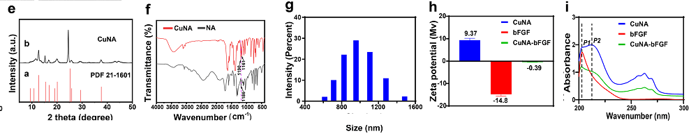
1d Powder X-ray diffraction spectra (XRD) showed that the CuNA peaks at 12.7, 15.28, 16.7, 20.02, 24.52, 25.88, 29.16 and 37.44° with little deviations, which are claimed to be assigned to a standard in 1e that we ill need to assume is PDF 21-1601.
Moving on to more physical characterizations:
Fig. 1 Characterization of CuNA prepared in ethylene glycol. (. e The X‑ray diffraction (XRD) patterns of CuNA. f FTIR of nicotinic acid and CuNA. g The size distribution of CuNA in distilled water. h Zeta potential of bFGF, CuNA and CuNA‑bFGF. i UV–Vis of bFGF, CuNA and CuNA‑bFGF
1e a standard…which can be assigned to the standards (JCPDS card no.
1f Peaks were assigned to C–O (COO–) stretching that suggests “strongly” that Cu is interacting with NA. More peaks were assigned to Cu interacting with the carboxyl group of NA rather than the nitrogen.
1g The dynamic light scattering gave an estimate of the particle size with an average diameter of ~ 980.8 nm,
1h Zeta-potential of CuNA is approximately +9.37 mV. Then, the Zeta-potential value of CuNA increases to −0.39 mV after loading with bFGF. Not shown on this post are zeta potential data showing that the addition of bFGF causes the particles to go from a negative to positive charge indicating that the bFGF had been loaded onto CuNA by a facile electrostatic adsorption.
1i In the spectrum of bFGF, the only absorption peak at ~ 202 nm is observed, which is all a little strange. Aromatic residues tend to absorb at 280nm. They were not looking at parts of teh visible spectrum at which Cu(II) and Cu(I) absorb.
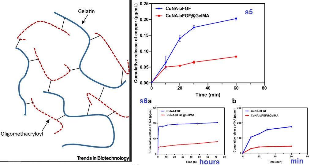
Gelatin methacrylate hydrogels
Wet GelMA and CuNA-bFGF@GelMA samples were frozen at - 80 °C and then lyophilized. The structure details and elemental mapping of GelMA and CuNA-bFGF@GelMA hydrogels were imaged by SEM. The surface chemistry of composite hydrogel was analyzed via XPS (Thermo Scientific, USA)
Figure S3 SEM and elemental mapping images of CuNA-bFGF@GelMA. Scar bar is 200 μm.
Not shown are supplemental Figure 3 X-ray photoelectron spectroscopy XPS results showing two populations of Cu: ~952 and ~932 eV. What these two populations represent was not discussed.
2. Loading gels and release of contents
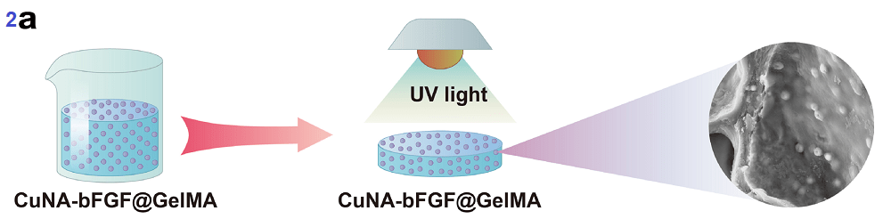
Figure 2 describes release of contents in hydrogels. What is perplexing is how these things were made. The gelatin methyl acrylate material was dissolved in PBS and heated to 37°C. Then the powders of CuNA or CuNA-bFGF were mixed with the solution at concentration of 0, 3, 5, 10 and 20 wt%. This material was poured into a Teflon mold and exposed to UV light and then stored at 4 °C overnight. We could probably modify this recipe by use of unflavored gelatin. The Cu(I)NA2 could be added as the mixture cools.
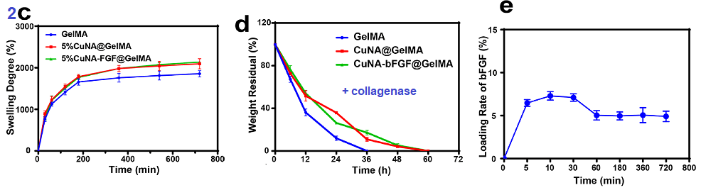
Figure s3 makes a lot more sense knowing what the structure of GelMA structures look like. It is interesting to note that a lot Cu is released from the GelMA structure than with just the bFGF. Even after about three days (70 hours, panel s6a)
Figure S5 The release profile of copper from CuNA-bFGF and composite hydrogel at first hour.Figure S6 The release profile of NA from CuNA-bFGF and composite hydrogel (a). The release profile of NA of 72 h; (b). The release profile of NA at first hour.
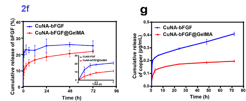
2c The swelling rate is proposed to play an important role in wound healing by absorbing wound exudates.
2d Type II collagenase was used in this study. Degradation of the gel is proposed to increase the rate of release of Cu and bFGF.
2e This figure refers back to adding bFGF to the Cu(II)NA crystals.
Both Cu(II)NA-bFGF crystals and the gel are slow release
If we wanted to do something sooner rather than later with Cu(I)NA2 and growth factors, we have reason to thing the release would be over the course of several days.
2f Gelatin-MA slows the release of basic fibroblast growth factor. The inset makes it clear that bFGF is release in an hour.
2g Mitosynergy already sells a Cu(I)NA2 cream. Might release be slower if collagen were added?
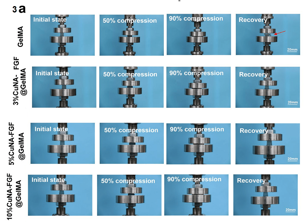
3. Mechanics of GelMA composites
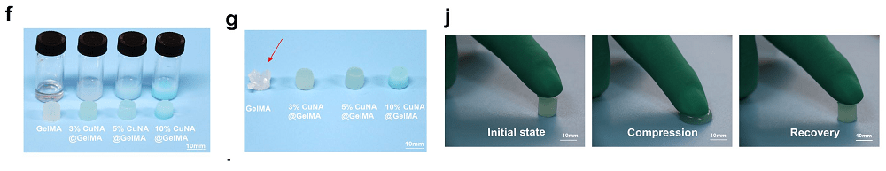
These are some lay person friendly images, from Figure 3 of the publication, that do not require the understanding of any hard core physics. Note that the CuNA composite is turquoise and clearly in the +2 oxidation state. We could probably make our own composite with gelatin and Cu(I)NA2 for visuals on how to increase the elasticity of one’s skin.
If 3g the arrow points to a gelatin-methylacrylate gel that cracked under the same stain that had no efect on the gesl with added caper.
Truly, the only thing in this study for us is the demonstration that what appears to be Cu(II)NA affects the mechanical properties of something that is likely to resemble collagen in our skin. Gelatin is essentially collagen. Figure 3 of the Wang publication is divided up into pieces to make it more comprehensible.
Figure 3a. Do we really want to invest the sort of resources required to replicate the results with a matrix of our choice and Cu(I)NA2?
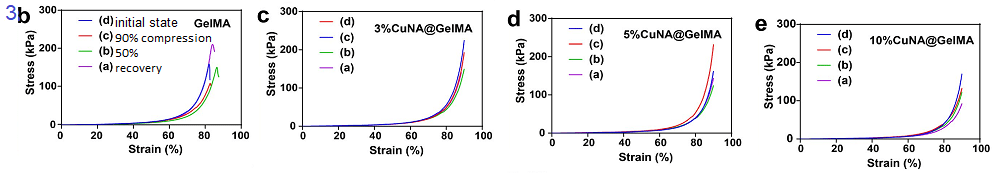
The above device is a universal machine tester (HY-940FS, Shanghai, China). The samples for the compression tests were prepared in 10 mm inner diameter cylinder molds. Compressive tests were conducted at a rate of 5 mm/min to the strain of 90%. Compressive young’s modulus was determined from the slope of the strain-stress curve between 10 and 15% strain. The cyclic compression tests were performed with ten cycles up to 50% strain followed the speed of 5 mm/min to characterize the mechanical properties of GelMA composites. Each sample was tested in quadruplicate. Moving on to panels 3b-f we are forced to consider the definitions of stress and strain. Stress is the force per unit area. Strain is the % change in length in this case. That the authors are measuring cyclical strain is the only way that these graphs can make sense.
Probably the best way of looking at these graphs is look at the x-axis and find the 80% mark.
3b the gel without added Cu take the less force (80kPa) to deform 80% of 50% deformed than when they at 90% of full length (100 kPa).
3c Adding 5% Cu(I)NA makes the gell easier to deform to 80% of whatever thickness it happens to be. and so on as the concentration of Cu(II)NA is increased.
Note that the authors did not look at bFGF.
The authors interpret all of the data as evidence that Cu(II)NA increases the elasticity of gelatin/ MA gels.
3h An example of the cycles of force applied to the gel. What they did not discuss that the viscous component is in phase with the cyclic force whereas the elactic component causes a resistance to deformation which lags behind the applied force. In other words, the gel bouncing back counters the force to compress again, hence elasticity.
3i Overall, less force (stress) is required to deform a gel 40% in which the copper has been added, even though it bounces back.
3k The compressive moduli calculated from the linear slope of the strain–stress curve shows decreasing values for GelMA, 3%, 5 and 10% CuNA@GelMAs respectively.
The authors claimed that ionic cross-linking effect between copper ion and GelMA molecule enhances the structure of composite.
There are those that tell you that mixing copper sulfate with ascorbate/vitamin C is the same as CopperOne Niacin. They are both a beautiful orange/red color, are they not? The are not the same! They are two perfectly and required nutrients that do not belong together. Just like….
For an explanation of what happens when you stick a fork into an outlet can be found on youtube.com.
Mr Fork and Mr Oulet can’t be friends because when you get the two together electrons go where they are not supposed to go. This is the same reason why Mr Copper and Mr Ascorbate cannot be friends. Getting the two together and a source of electrons can result in those electrons going where they shouldn’t.
A silly animate and dialog associated with some equations explaining why copper and Viatamin C (ascorbate) are pro-oxidants
These are the sources of the animation
Mastrangelo D, Massai L. Vitamin C against Cancer. , Chapter 4 in VITAMIN C Edited by Amal H. Hamza Published by InTech Janeza Trdine 9, 51000 Rijeka, Croatia This book chapter was used for equations 1-4.
Belmonte M, Elhiti M, Waldner B, Stasolla C. Depletion of cellular brassinolide decreases embryo production and disrupts the architecture of the apical meristems in Brassica napus microspore-derived embryos. J Exp Bot. 2010 Jun;61(10):2779-94. PMC free articleThis article was used to fine tune the relationship between ascorbate free radicals and the anti-oxidant small molecule glutathione, GSH
Vitamin C and Fenton reaction [1]
Note that Cu2+ can be substituted for Fe3+ [2] Radicals, unpaired electrons, in these equation are denoted by superscript”â—“
Fe3+ + AscH2 → Fe2+ + Ascâ—- → + 2H+
Fe2+ + O2 → Fe3+ + O2â—-
2O2â—- + 2H+→ H2O2 +O2
H2O2 + Fe2+ → Fe3+ + OH– + OHâ—-
The Letelier study started with the observation that copper ions can irreversibly and non-specifically bind to thiol groups in proteins. They noted that this non-specific binding property was not as fully addressed for iron ions.
Cu2+/ascorbate elicited more oxygen consumption than Fe3+/ascorbate under protein free conditions. [2] see equation #2
In the presence of cytosolic and microsomal protein, Cu2+/ascorbate increased microsomal lipid peroxidation and decreased cytosolic and microsomal content of thiol groups more efficiently than Fe3+/ascorbate. [2]
Finally, Fe3+/ascorbate and Cu2+/ascorbate inhibited in different ways cytosolic and microsomal glutathione S-transferase (GST) activities, which have subtle differences in sensitivity to oxidants.
In the absence of ascorbate, only Cu2+ decreased the content of cytosolic and microsomal thiol groups and inhibited cytosolic and microsomal GST activities.
The reaction
This reaction was pieced together from references [2] and [3]
Ascâ—- → dehydro Asc +e–
dehydro Asc + 2GSH → Asc + GSSG
Now that the Ascorbate has been regenerated it is free to start the reactive oxygen species regeneration again with more Cu2+.
So why can’t Copper and Vitamin C be “friends”?
Because when the two get together, electrons go where they shouldn’t be.
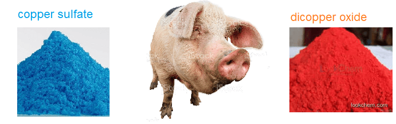
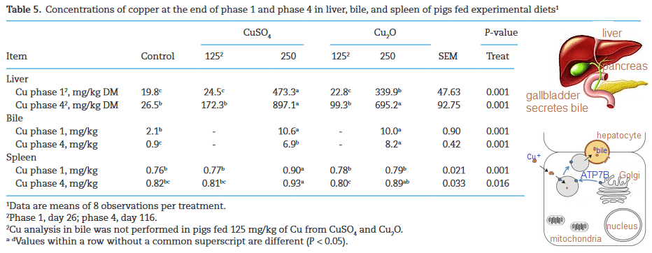
The Blavi 2021 study demonstrated that dicopper oxide is a superior to the common copper sulfate supplement in pigs fed the enzyme phytase to make plant phosphate more bioavailable.
We at CopperOne would like to say that the leading dietary copper supplement, cupric sulfate (CuSO4), was compared side by side with cuprous nicotinic acid and the latter was found to be superior in a large species that resembles humans. This is dimply not the case. A team from France, Spain, and the United states compared cuprous oxde (C2O) with CuSO4 in the feed of finishing pigs. Our original thought was that these experiments were conducted because the authors considered cuprous copper more bioavailable. The concern was that Cu2+ is an inhibitor of an enzyme called phytase, which which makes phosphate found in the plant compound phytic acid more bio available. The featured image came from Bacillus amyloliquefaciens. Pytase binds Ca2+ as part of the process of removing phosphates from the plant compound phytic acid. Since the enzyme phytase is a widely used feed supplement to increase the bio availability of phosphate of plant feed in farm animals, Blavi and coauthors are proposing that Cu2+ supplements may affect bone mineralization by preventing the absorption of phosphate from phytic acid.. Their concerns were also centered around Cu2+ binding to phytic acid more so than Cu2+ binding to Ca2+ sites on supplemental phytase.
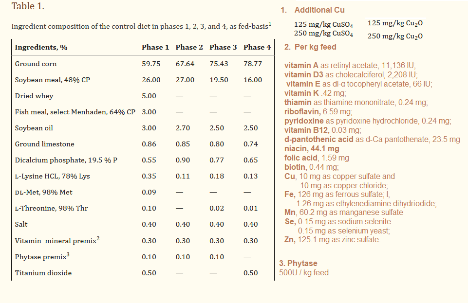
1. The Diet
As pigs grow, their nutritional needs change. This study used slightly different feeding schemes for different stages of growth. At the end of each stage, a representative pig of the group was slaughtered and analyzed for bone mineral content and other parameters.
Phase 1, days 1 to 26
phase 2, days 26 to 61
phase 3, days 61 to 96
phase 4, days 96 to 116.
Note that the pigs are already being supplemented with niacin. In an actual farm setting, why not use cuprous nicotinic acid instead of di cuprous oxide?
2. The diets
The take home message is that the diets are pretty similar.
3. Weight gain
The 250 mg/kg Cu2O supplemented pigs (superscript “a”)gained more weight in all four phases than the controls(superscripts “b” and “c”) . Pigs on the 250 Cu2O gained more weight in phases 3 and 4 than their 250 CuSO4 counter parts.
Table 2 from Blavi 2021 with a cartoon pig pointing to the significant difference between the control and the equivalent about of copper sulfate in phases 3 and 4.
Perhaps the other main point of Table 2 is that the pigs on Cu2O were not eating more than the pigs on the CuSO4.
4. Other meat industry parameters, no difference
Note that these pigs already had supplemental copper in their diets by way of the mineral/vitamin mix in Table 1. Additional copper made no difference in these parameters.
5. 250mg/kg Cu is too much?
This may not have been what the authors were considering at first, but look at the numbers. Going from 125 to 250 mg/kg Cu in the diet increases copper in the liver over 10x in Phase 1. In Phase 4 the increase is not as dramatic. Less copper accumulates in the liver in the 250 Cu2O group. Export in the bile is naturally much greater than in the control group. By Phase 4 more Cu+ was being exported in the bile in the Cu2O group than in the CuSO4 group. Was this because the ATP7B export pump uses Cu+. We can only speculate at this point.Mutations in ATP7B define Wilson’s Disease, which is defined by copper poisoning by failure to export excess in the bile.
Why the spleen was examined was not entirely clear. The spleen may play a role in the immune system and processing of damaged red blood cells, but its role in copper handling is less clear. Perhaps the spleen was simply in the abdominal cavity neighborhood. At the potentially toxic copper concentration of 250mg/kg, the copper content of the spleen was statistically the same as the control in the 250 Cu2O group but elevated in the CuSO4 group.
6 Bone mineralization
Table 6 has been divided into two sections just because there are two points to be made by the information by this table. Recall our first issue with CuSO4 is that it might bind to phytase and decrease the pigs’ ability to make use of phosphate bound to phytic acid in the soy and corn they were consuming. Bone is composed of collagen fibers with hydroxyapatite deposits. Hydroxyapatite is calcium and phosphate. No changes were seen in the percentages of calcium and phosphate in the bone ash. One would expect that burning the bone to ash would remove all traces of collagen.
Table 6 from Blavi 2021 edited to only show calcium and phosphate data.
The total amount of noncombustible (ash) in the bone is greatest in the 250 Cu2O treatment. Calcium data is a bit more ambiguous. We can say that 250 Cu2O is better than 250 CuSO4 but we cannot say Cu2O is better than the control diet with no additional copper. The bone phosphate data confirm the investigators’ concern that CuSO4 is inhibiting phytase. 250 Cu2O treated pigs have (statistically) the same phosphate content in their bones as the control group of pigs.
Moving on to the copper and zinc component of Figure 6 things get more complicated and interesting. It is assumed mg/kg means mg/kg bone ash and that mg/kg means mg/kg non combusted bone.
Note that both copper compounds decrease the amount of zinc (in bone ash?) in Phase 1 but not Phase 4.
What did the authors discuss and conclude?
They mentioned that Cu is absorbed by the intestine in the +1 oxidation state rather than the +2 state. They discussed Cu2O being less water soluble than CuSO4, less likely to dissolve, dissociate into ions, and form complexes with phytic acid. The intestinal pH was also discussed somewhat. The authors also discussed some previous studies that we may look into some more. Their review of the literature led them to conclude that hepatic copper is a good indicator of copper absorbed in farm animals. We at CopperOne would probably be speculating too much if we proposed Cu2+ could get stuck in the liver and never transported out of the liver on ceruloplasmin. Certainly more copper got incorporated into the spleen in CuSO4 fed pigs, see figure 5. This is a very interesting study that has given us a lot to think about.
Wikipedia has a good page on AL amyloidosis. “AL” stands for amyloid light chain. The light chains are part of the antibody molecule. AL is linked to diseases associated with increased antibody production: B cell myeloma and Waldenström’s macroglobulinemia. The Wikipedia authors did not mention of the therapeutic antibodies being injected into patients to treat what seems like every conceivable disease. We can only hope that these therapeutic antibodies are fully assembled and do not contain rogue light chains that can form amyloids and other toxic compounds with Cu(II)
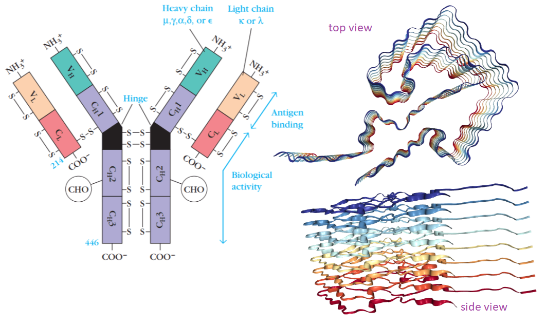
What is an amyloid anyway? The featured image comes from a structure deposited on RCSB.org, a public access database of protein structures. The image of the IgG antibody molecule was obtained from this link. Note the presence of disulfide bonds (S-S) in the light chains. Also note that the light chain has a constant CL and a variable VL region. For readers that like to attribute human characteristics to molecules, it is as if the misfolded deep red light chain says to a red/orange light chain buddy in the process of folding and getting attached to a heavy chain, “Why do you want to conform when you can be deviant like me?” Deep Red and Red Orange spoon together in the same deviant pattern. Then the deviant pair say to the orange light chain in line to be made into an antibody, “Why are you conforming when you can be deviant?” The deviant trio recruit the Bright Yellow light chain into their pack of biological aberration. The pack grows and grows until they become large enough to get trapped in places where they can cause trouble. The heart, of course the heart! The reader is invited to go to the RCSB.org, website and rotate the structure around to get a feel for what an amyloid really is. The curious thing is that this structure is based on a fibril isolated from an actual patient. [1] How scary is that?
Basics of the amyloid structure
Radamaker and colleagues from several universities in Germany isolated AL amyloid fibrils from the heart of a woman suffering from advanced heart failure due to AL amyloidosis. [1] The patient was diagnosed with a monoclonal plasma cell disorder called “smoldering myeloma” one year before she was diagnosed at the same time as AL amyloidosis. The patient was treated for the bone marrow plasma cell cancer but required a heart transplantation anyway due to amyloid deposit build up in her heart. [1] The authors used an established protocol to isolate the amyoid fibrils from her diseased heart. [1]
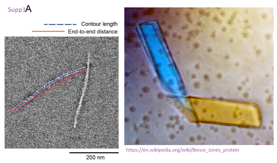
The authors used cryoelectron microscopy to predict the structure of her light chain amyloid deposits. CryoEM involves taking a transmission electron microscopy image of a structure and fitting in fitting in known structures of the protein, or related proteins, based on X-ray crystallography data. In this case the scientist used a combination of the sequence of her light chain and the 1BJM entry of the rcsb.org database. To be specific, the X-ray crystal structure was based on Bence-Jones crystals of light chains found in the urine of multiple myeloma patients.
Supplemental figure 1A from Radamker2019 showng a transmission electron microscope image of an AL fibril. also shown is are some Bence-Jones crystals of IgG light chains from Wikipedia.
Okay, now that we are dealing with IgG light chains behaving badly, let us get on to our story of how Cu2+ makes a bad situation worse. This sequel comes from collaborators from Italy. [2]
Cu(II) / Cu2+ making a bad situation worse
Histidine is generally considered an amino acid that likes to bind to Cu with the imidizole nitrogen shown in blue below. Cysteine has a thiol that also likes to bind Cu, but in our case they can’t because they are bound to each other in a disulfide bond.
The Cu2+ sites in relation to structure. A. sequence and structural information of the amyloid from RCSB.org, The golden bars are beta sheets, the structures that sort of look like folded bed sheets in the featured image. The disulfide bond cysteines are shown for reference. B. The sequence of amyloid forming light chain from Russo 2022 [2] These authors mutated the Cu2+ binding sites His188 and His197 to alanines. The magenta paint brush sweeps represent other amino acids of the IgG light chain in the vicinity of the histidines being mutated to alanines.
Only the side chains are shown just to make the point that a lot of Cu2+ binding groups are lost in going from histidine to alanine with a side chain consisting of one carbon with three hydrogens that chemists don’t generally draw unless they can come off.
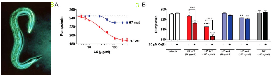
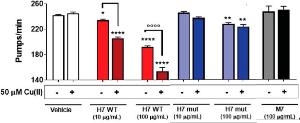
Most lay readers will want to know about toxicity. Rosso and coworkers used a nematode called Caenorhabditis elegans or C elegans for short. They have an ancestral heart and circulatory system that can be observed to “beat.” These wormies were fed eh IgG light chain proteins with and without small amounts of Cu2+. Note that the wild type H7 light chain becomes more toxic with 50 µM Cu2+. [2] In panel 3B, M7 are non amyloid genic IgG light chains from multiple myeloma patients that just happen to be non amyloid genic.
This is the link to the image on the left. H7 mutant with destabilized CL domain is less toxic to C. elegans. (A) Dose–response curves reveal the diminished toxicity of H7-H188A/H197A (H7 mut) compared to H7 (WT), as higher concentrations are required to inhibit the pumping rate of worms. Worms were fed for 2 h with different concentrations of WT or mutated H7 suspended in 10 mM PBS, pH 7.4, and the pharyngeal pumping was scored 24 h after the administration. Control worms received vehicle alone (dotted line). Each value is the mean ± SE, n = 30. IC50 was 28.9 and 46.8 µg/mL for WT and mutated H7, respectively, p < 0.01, Student’s t-test. (B) Worms were fed for 2 h with 10 or 100 µg/mL H7 or H7-H188A/H197A, or 100 µg/mL M7 dissolved in 10 mM PBS, pH 7.4, with or without 50 µM Cu (II). Control worms received 10 mM PBS, pH 7.4 with or without 50 µM Cu (II) (vehicle). Pharyngeal pumping was determined 24 h after the administration. Each value is the mean ± SE, n = 20. * p < 0.05, ** p < 0.01 and **** p < 0.001 vs. the corresponding vehicle, °°°° p < 0.001, one-way ANOVA and Bonferroni’s post hoc test.A close up to show just how toxin Cu(II) is in the presence of IgG light chains!
This post is going to skip some very complicated biophysical techniques that tested thermo stability of the amyloidgenic light chains. [2] Data not shown demonstrate Cu2+ binding to the VL of the cardiotropic H7 with a low micromolar affinity. [2] Alanine-substitution of two histidine residues in the constant domain of H7 (H7-H188A/H197A mutant) did not alter Cu2+ binding or impair H2O2 generation in vitro. [2] The His-to-Ala mutations destabilized the constant domain and reduced the toxicity of the H7 mutant. Rosso and coauthors could not explain the toxicity of Cu2+ only to the His188/His197 binding site alone. [2]
Two things to look into, in my opinion, would be
Cys221 at the end of the sequence and whether or not the disulfide bond was really there.
Free -SH groups, as opposed to -S-S- in disulfide bonds, could bind Cu2+.
This is an exciting paper in so many ways.
What if we were to take a casein digest mix of peptides, add a small amount of Cu2+ or Cu+ and feed them to C elegans? A next step could also be to use florescent dyes that bind to amyloid structures. Thioflavin T is just one example.
References
Radamaker L, Lin YH, Annamalai K, Huhn S, Hegenbart U, Schönland SO, Fritz G, Schmidt M, Fändrich M. (2019) Cryo-EM structure of a light chain-derived amyloid fibril from a patient with systemic AL amyloidosis. Nat Commun. 2019 Mar 20;10(1):1103. PMC free article
Russo R, Romeo M, Schulte T, Maritan M, Oberti L, Barzago MM, Barbiroli A, Pappone C, Anastasia L, Palladini G, Diomede L, Ricagno S. Cu(II) Binding Increases the Soluble Toxicity of Amyloidogenic Light Chains. Int J Mol Sci. 2022 Jan 16;23(2):950. PMC free article
This post is not intended to make any medical claims to treat Long Covid, Post Treatment Lyme Disease, or any other post infection chronic condition. In a previous post different tests were reviewed to The maker of CopperOne would like to conduct clinical trials so that such claims could legitimately be made. Questionnaires are great. They minimize the use of needles for blood draws. Redman and coworkers administered many neurological questionnaires. [1] This test was published in a journal that seems to have died out. http://orthomolecular.org/library/jom/index.shtml
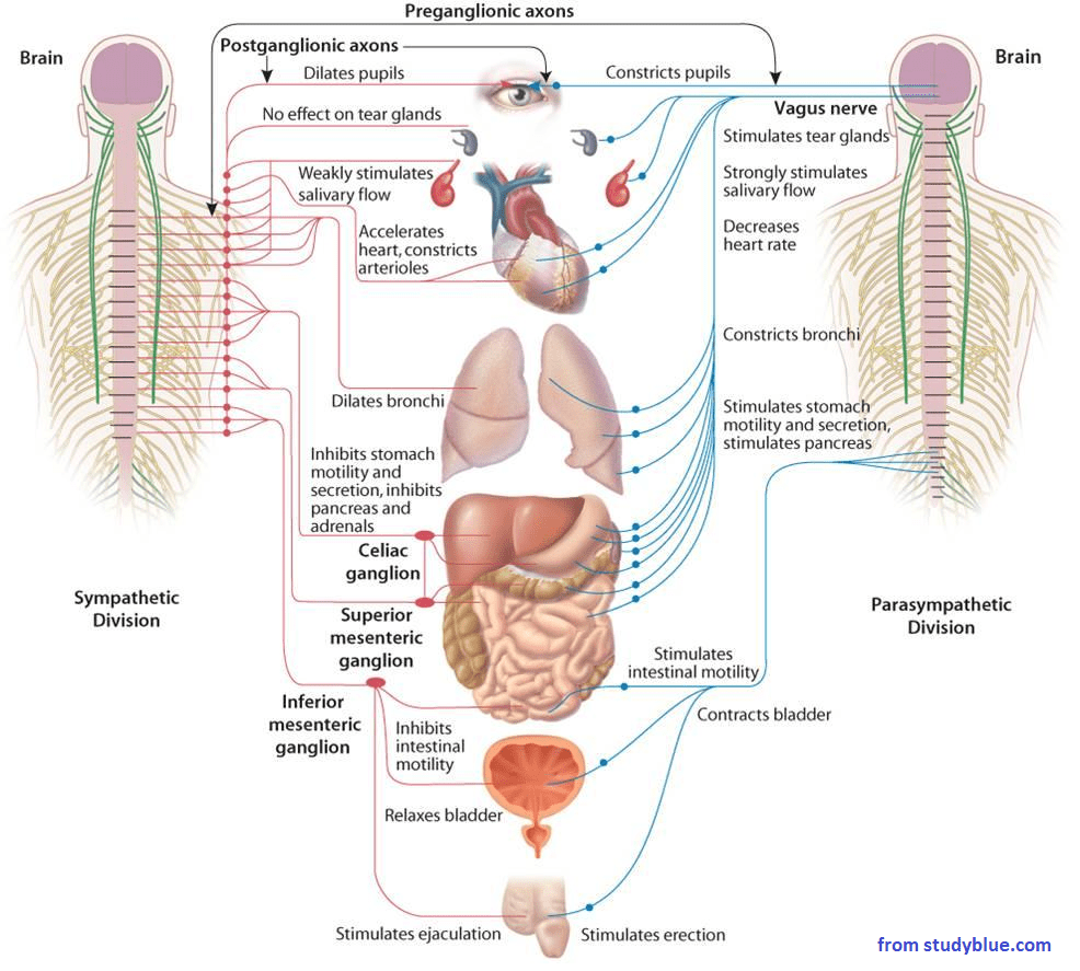
The trouble with our last clinical trial is that the participants may have gotten bored answering the two questionnaires and and might have checked boxes without thinking. This gem published back in 1977 seems to be more interesting and to the point. The literature on Covid-19 and the autonomic nervous system is impressive. Dividing symptoms between sympathetic and parasympathetic branches is less common. We have covered how Long Covid symptoms are extremely similar to post treatment Lyme Disease (PTLD).
As a Long Covid or PTLD patient, do you see yourself in these questions? Does the switch from the cholinergic to the adrenergic questions force you to pay attention?
symptom
15 points
10 points
5 points
0 points
symptom
very frequently
often
sometimes
rare or never
diarrhea
heartburn
urge to frequent urination
one or more muscle feel weak
sleeping more than usual
flushing of the face
waterish mouth
excessive appetite
feel sexually aroused
have difficulty breathing
dry palms
Begin Cholinergic
constipation
one or more muscles feel tense
sleeping less than usual
palor of face
cold feet
wet hands
we armpits
heart palpitations
dry mouth
goose pimples
butterflies in stomach
poor appetite
From reference [2] Frequency of the symptoms were given the following values: very often —15; often —10; sometimes—5; and rare or never—0.
If you already are a CopperOne customer, did the answers to any of these questions change when you started?
Referees
Rebman AW, Bechtold KT, Yang T, et al. . The clinical, symptom, and quality-of-life characterization of a well-defined group of patients with posttreatment Lyme disease syndrome. Front. Med. 2017;4:224 10.3389/fmed.2017.00224 [PMC free article]
Neziroglul F and Yaryura-Tobias J A (1977) Development of an Autonomic Nervous System Questionnaire: Diagnostic Aid in Measurement ofAnxiety, Depression, and Aggression. Orthomolecular Psychiatry vol 6, no 3, 1977, Pp. 265-271 free article
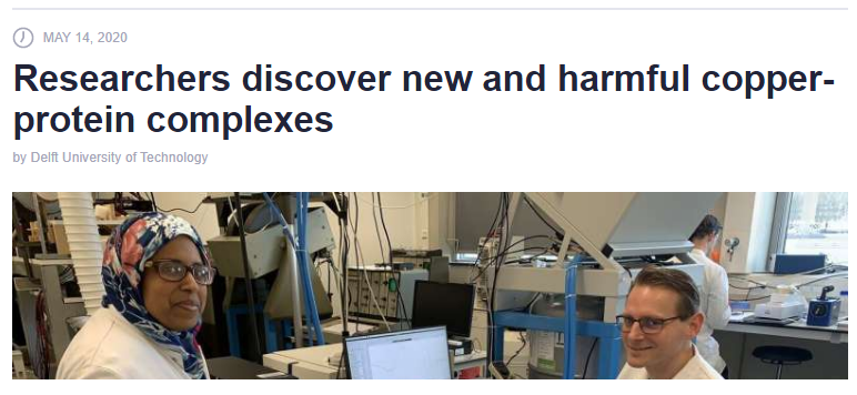
Many of us may be accustomed to getting suspicious sounding science news on our cell phones. A customer sent Mitosynergy this link .  Sure enough, I got the same story on my cheap smart phone. Fake News!! Unfortunately the new clip did not do a very interesting study justice. The author did provide a link to the online publication.
Summary/abstract for Scientists
“The aminoâ€terminal copper and nickel/Nâ€terminal site (ATCUN/NTS) present in proteins and bioactive peptides exhibits high affinity towards CuII ions and have been implicated in human copper physiology….One of these novel intermediates, characterized by twoâ€nitrogen coordination, t1/2 ≈100 ms at pH 6.0 and the ability to maintain the CuII/CuI redox pair is the best candidate for the longâ€sought reactive species in extracellular copper transport.”
Why I think this is a cool paper for Mitosynergy and its customers
Charlie Barker asked me if one could see CuII/CuI redox cycling color changes of Cu in cytochrome C oxidase in the mitochondria. My reply was that if you could build a spectrophotometer small enough one could.
Charlie Barker made the big deal that oxidation and reduction are dependent on pH. This is true.  This is what charged amino acid side chains do in enzymes. They control the local pH, never you mind what the pH is in the bulk environment.
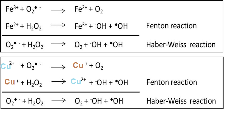
This is the problem with these CuII/CuI redox cycling peptides. There is absolutely no local pH control. There is absolutely no control of where they may pick up electrons Shown there are Fenton and Haber Weiss reactions. Hydrogen peroxide and superoxide may be products of the mitochondria.
Some redox cycling reactions involving iron and copper
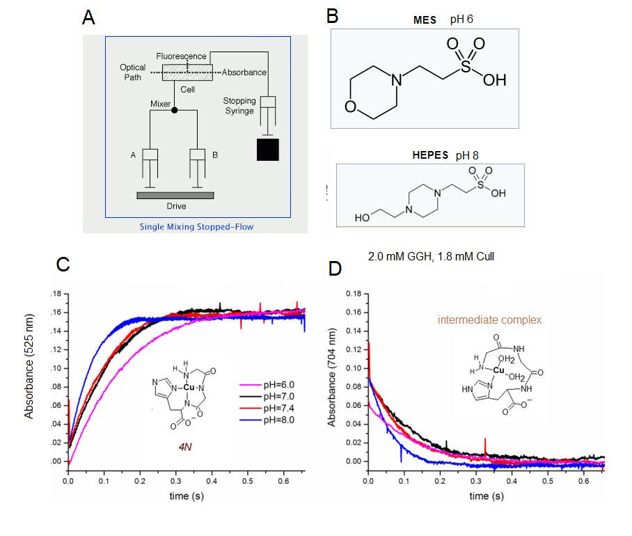
What is stopped flow?Â
Stopped flow is a way of measuring the kinetics of a reaction. I’ve taken much liberty in embellishing supplemental figures so that you, the lay reader, can understand this very interesting work. In panel A we have the basic stopped flow design. The one I used in grad school had a trigger that released compressed air on the drive that pushed a set amount of contents in syringe A and syringe B and sent them to the mixer. The same trigger that released the compressed air also started measuring fluorescence in the optical cell. The same trigger that released the compressed air also started the recording. of the reaction going on in the optical cell. These authors measured absorbance. In Panel B I added the structure of the buffers they used to set the pH of the reactions. Note that MES and HEPES have very similar structures.  These authors are careful and know their stuff.
What is stopped flow A. The schematic of a stopped flow spectrophotometer. B The authors controlled he pH of the reaction with similar buffers C. As the product of the reaction increases, so does its absorbance at 525 nm. D. As an intermediate decreases, so does its absorbance at 704 nm.
In panel C we have a time dependent increase in the absorbance of what Kotuniak concluded to be the final product, 4N. Naturally, the absorbance of the intermediate decreased with time. Note that all of this happens in less than a second.  If our eyes were fast enough, we’d see a cyan colored complex turn to a sort of magenta color, based on its absorbance of green light. Note that the higher the pH, the faster the reaction with Cu2+. No reaction was seen at stomach pH. If this reaction occurs in humans, it is as peptides and Cu2+ enter the duodenum.  Now that we’ve covered what stopped flow is, now we need to cover what a peptide is.
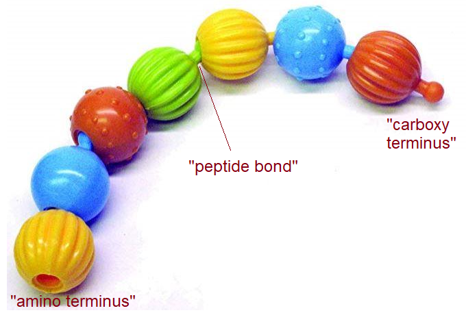
Some peptide basics
Amino acids link together to form peptides and proteins just like pop beads link together to form strings. The “carboxy terminus” of one bead fits into the “amino terminus” of another bead, and so on.Peptides are made up of amino acids. Amino acids have an NH2 group on the amino terminus and a -COOH group on the carboxy terminus. By abstracting an H20 a “peptide bond” is formed.
Amino acids link together to form peptides and proteins just like pop beads link together to form strings. The “carboxy terminus” of one bead fits into the “amino terminus” of another bead, and so on.
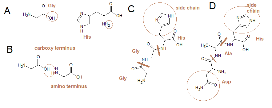
What is this  aminoâ€terminal copper and nickel/Nâ€terminal site (ATCUN/NTS) ?
We now know what a peptide bond and N-terminus are. The Cu/Ni site is any two N-terminal amino acids and a histine in the number 3 position. The N-terminal sequence woudl be X-X-His where X = any amino acid.  The authors used Glycine because it is the simplest amino acid. Panel A shows the structures of the amino acids Glycine and Histidine. The carboxy terminus of Gycine and hte amino terminus of Histidine have been circled. Panel B shows the formation of a peptide bond between two glycines Panel C shows the structure of the Gly-Gly-His peptide. Heavy lines denote the peptide bond. The histidine side chain is circled panel D.
A Structures of the amino acids Glycine and Histidine. B. Formation of a peptide bond between two glycines C. Structure of the Gly-Gly-Hist peptide. Heavy lines denote the peptide bond. The histidine side chain is circled. D. The same motif motif from human serum albumin.
The authors mentioned human serum albumin in their review of the literature.Let’s take a closer look and see what they mean.
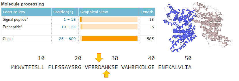
Here we have an image of a human albumin dimer along with the first 50 amino acids of its sequence. The signal sequence directs the growing protein to the Golgi for secretion into the blood. The pro-peptide is cleaves off again. The actual chain is whittled down to amino acids 25-609. This brings us down to the “toxic copper”binding site on the N-terminus.
Based on pH alone, we can expect this peptide to bind copper in the blood.
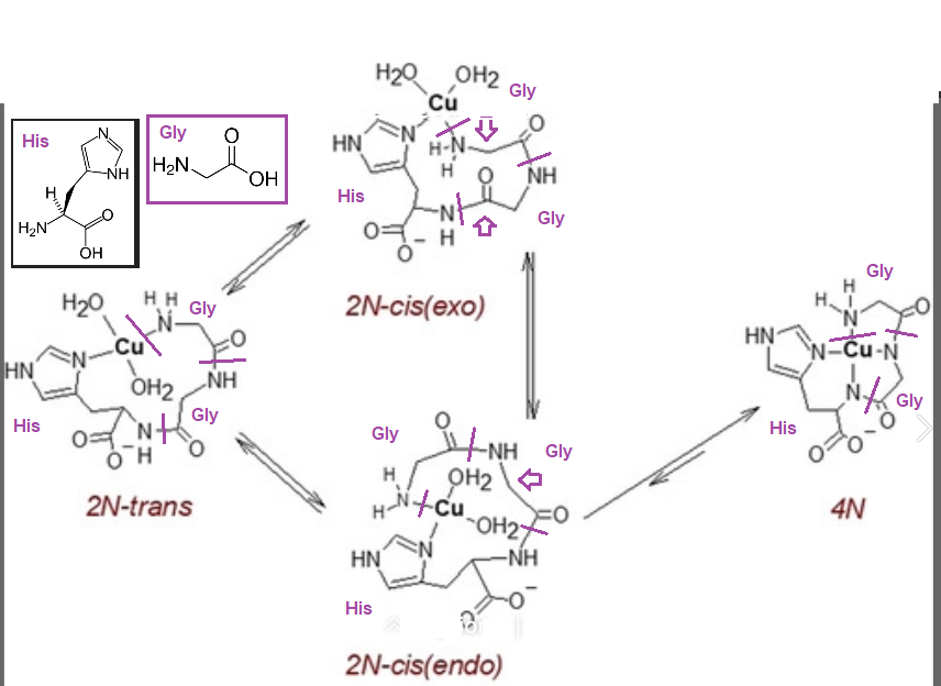
How does Cu2+  bind to the ATCUN/NTS motif?
We should add that Kotuniak were also able to freeze the reactions and use EPR to solve the structures of the reaction intermediates and product.
This is scheme 2 from the Kotuniak (2020) paper. The structures of glycine and histidine have been inserted in the upper left hand corner.  Dark, magenta lines have been drawn through the peptide bonds. Amino acids have been labeled. Thick, hollow, magenta arrows point to where changes are occurring.
And what about redox cycling?
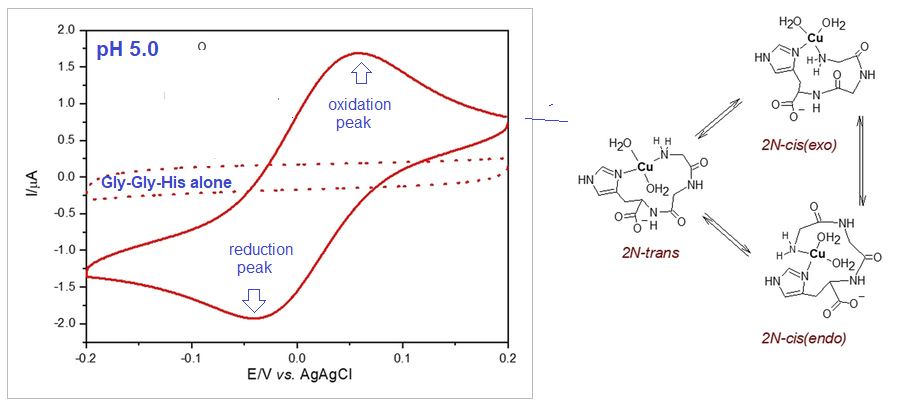
Kotuniak (2020) used cyclic voltametry to measure the redox potential of the complexes. Wikipedia gives a rather nice overview of this rather complicated physical technique.
Left, adapted from Figure 4 Kotuniak (2020) Right The cis-trans isomerization of the 2N complex was proposed to be the source of redox cycling
This brief overview is not giving the publication or the ample supplemental information that supports the publication. The authors referenced previous work on scanning voltammetry work performed on Azheimer’s Disease Aβ peptides.
Implications of this work for Mitosynergy
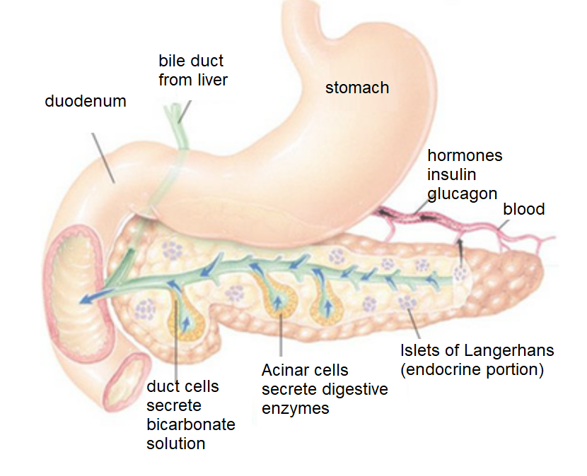
Kotuniak demonstrated that the copper complexes do not form at pH 1-3 of the stomach. As partially digested food leaves our stomach and enters our duodenum, our pancreas secretes bicarbonate and digestive enzymes.
Kotuniak (2020) were careful to use chemically related buffers. What if we were to repeat these experiments with physiological buffers?
We could have peptides and a standard Cu2+ supplement and peptides in one stopped flow syringe and a bicarbonate solution in another.
Does this “toxic copper” form complexes in duodenum conditions that can redox cycle?
What about Cu(I)NA2?
Can Cu(I)NA2 donate Cu+ to peptides?
Kotuniak R, Strampraad MJF, Bossak-Ahmad K, Wawrzyniak UE, Ufnalska I, Hagedoorn PL, Bal W.(2020)Key Intermediate Species Reveal the Copper(II)-Exchange Pathway in Biorelevant ATCUN/NTS Complexes. Angew Chem Int Ed Engl. 2020 Apr 8. doi: 10.1002/anie.202004264. [Cross Ref]