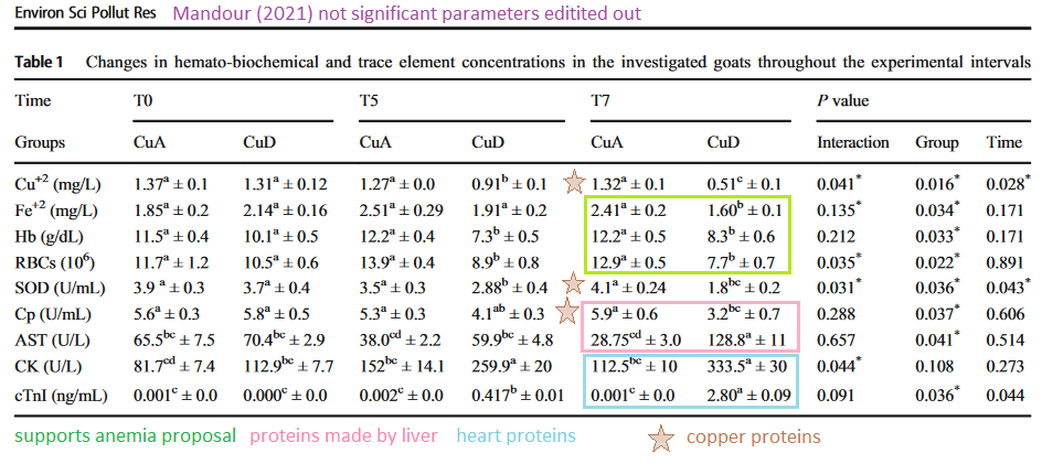
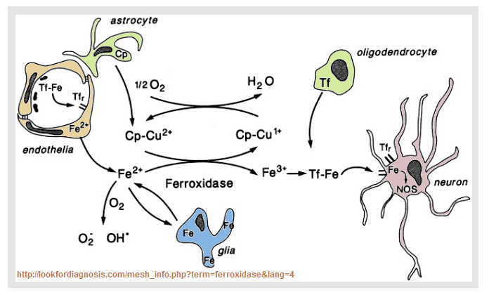
Copper is a heavy metal and is needed for myelin development. [1-3] Surely too much of a good thing is bad. Often other heavy metals follow copper. A study out of Rhode Island USA correlated neurological health with placental heavy metal concentration. [4] A study out of Taiwan looked at heavy metals and phtalates in the urine of pregnant mothers. [5] Both studies correlated heavy metals with mental functioning of the children. Phtalates are used in the production of plastics. When an expecting mother has concerns about heavy metals in the town’s water supply she drinks more bottled water right? This post makes a weak attempt to explain the statistics used in the Rhode Island and Taiwan studies. The take home is that prenatal copper is likely a good thing. Phthalates are probably bad. You really need to discuss the issue with someone licensed to practice medicine.
- Copper deficiency and myelin
- Copper excess a bad thing?
- Other sites on prenatal copper
- Talk to your healthcare provider
- References
Copper deficiency and myelin
Copper is essential to prenatal development of all organ systems. [1] Recently copper deficiency has become more prevalent due to zinc supplementation and bariatric surgery. [1] This same review claims that persistent structural changes can occur that supplementation after birth may not repair the damage. [1] In a 1976 rat study copper deficiency induced by a low copper diet for three generations of rats. [2] These authors demonstrated reductions in the yield of myelin (56%), brain weight (11%), and body weight (43%) in F2 generation rat pups nursed by their own copper-deficient mothers. [2] The myelin associated glycoprotein appeared to have a higher molecular weight. Normal myelin weight was restored when the pups were allowed to nurse from a healthy mother that was not copper deficient. [2] Brain and body growth were not restored. [2] This postnatal copper replacement by a foster mother produced a normal yield of myelin per gram of brain tissue, but.
Close to 50 years later Wikipedia authors have more to say about myelin associated glycoprotein MAG. The full length membrane associated protein is 100kDa, the cleaved portion is no long associated with the oligodendrocyte membrane. This image came from ResarchGate.
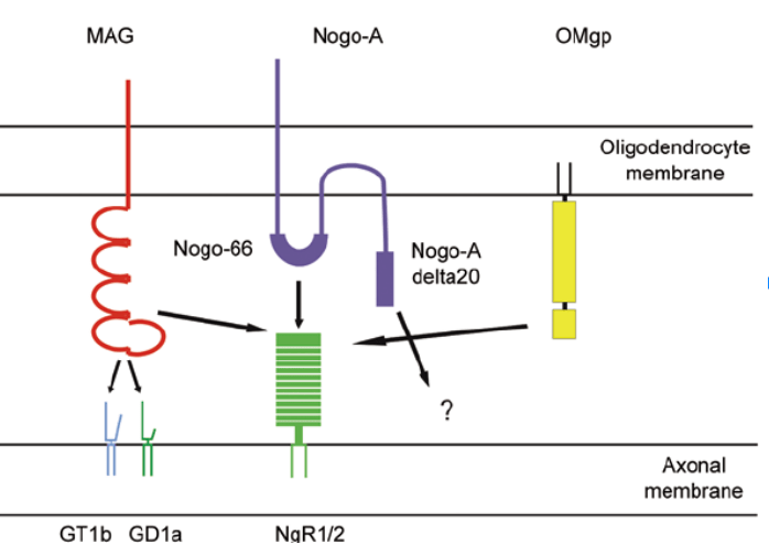
Myelin inhibitors of axon regeneration. Myelin-associated glycoprotein (MAG) halts axonal regrowth by binding to the gangliosides GD1a or GT1b and/or Nogo receptors 2 and 1 (NgR 2/1). Neurite outgrowth inhibitor A (Nogo-A) contains two axonal outgrowth inhibitory domains, Nogo-66 and Nogo-A delta20. The receptor for Nogo-66 is NgR1, while the receptor for Nogo-A delta20 is unknown. Oligodendrocyte myelin glycoprotein (OMgp) inhibits axonal regrowth by binding to NgR as well.
Lessons from the cuprizone model of MS
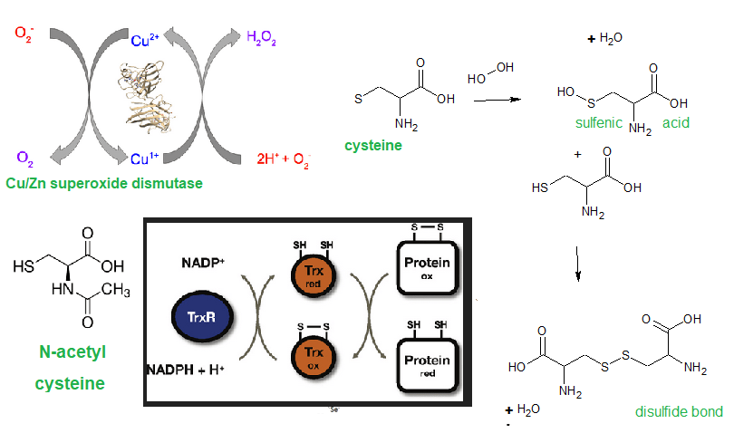
Cuprizone is a copper chelator that that is used to induce experimental models of the demyelinating disease multiple sclerosis. On one many peer reviewed studies from this PubMed search [3] points to the mitochondria and Cu/Zn SOD as being targets.
- Oligodendrocytes are the myelinating cells that increase in numbers during mid gestation. Copper deficiency leads to excess reactive oxygen species due to failure of Cu/Zn supreoxide dismuatase to scavenge and, secondly, rupture of mitochondia membrane, release of cytochrome C and apoptosis. [3] Cuprisone selectively decreases the mitochondria membrane potential of this cell type. [3]
- Astrocytes help form the blood brain barrier and are responsible for uptake of excess neurotrasmitter. A Cuprizone activation of astrocytes may contribute to demyelination. [3]
- Microglia are the macrophages of the brain. Cuprisone causes the accumulation of microglia and macrophage. [3]
We hear a lot about copper toxicity? What gives?
Copper excess a bad thing?

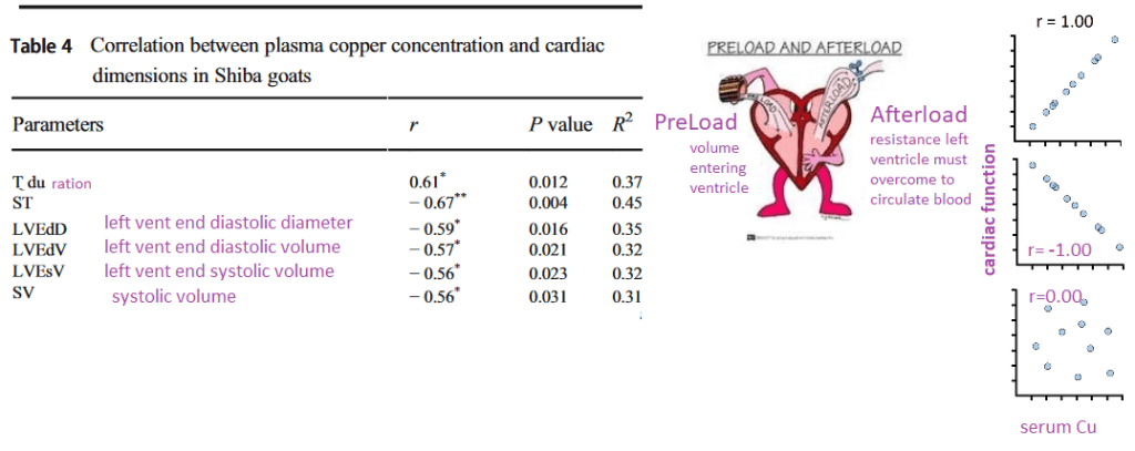
The Rhode Island study measured the heavy metal content of the placenta and assessed the infants soon after birth.[4] The Taiwanese study measured the heavy metal content of the mother’s urine during pregnancy and assessed the children as toddlers. [5]
The Rhode Island study suggests that low copper in the placenta is associated with worse category of neuro-behavior deficits. [4] The Taiwanese study suggests that high copper in the urine is associated with neurological issues in toddlers. [5]
In Rhode Island USA
Data from the Rhode Island Child Health Study (RICHS) population was used to test the hypothesis that placental heavy metals are associated with neurological problems. .NICU Network Neurobehavioral Scale (NNNS) was used to assess behavior issues in infants. [4]
Defining profile behaviors
Figure 1 breaks the study population children into a series of functional Z scores.
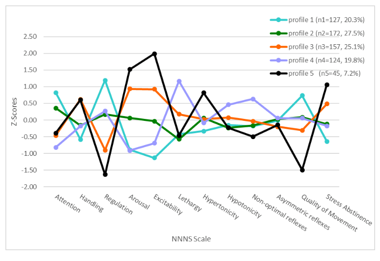
- highest attention, movement regulation, lower stress abstinence signs, less handling, excitability hypertonicity.
- typical neurobehavior, largest profile, overall average, except lowest in the lethargy scale.
- average performance of scales
- lethargy, hypotonicity, non-optimal reflexes, and lowest attention and arousal.
- most extreme regulation, arousal, excitability, hypertonicity scores, more non-optimal reflexes, lowest quality of movement and highest stress abstinence signs.
Heavy metal levels by profile groups
Note that these are placental heavy metal concentrations.
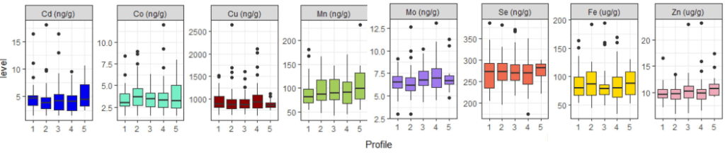
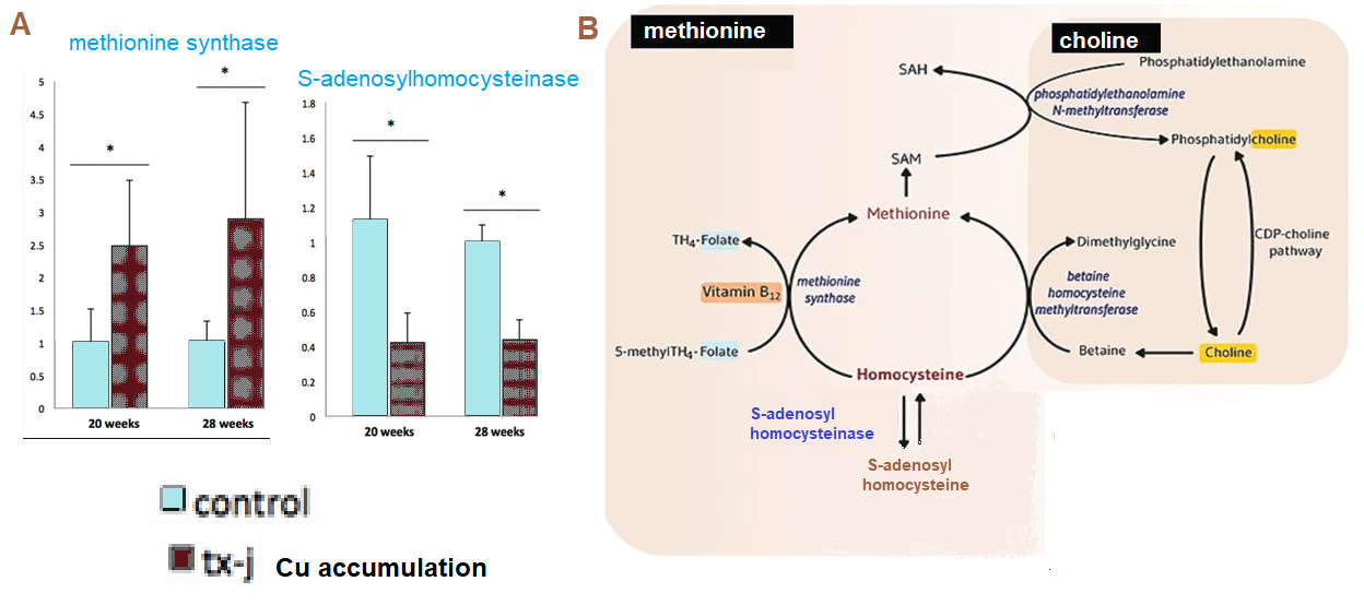
These box and whisker plots show median (line through the box) and the boundaries of the 2nd and 3rd quartiles. The lines mark the 1st and 4th quartiles. The dots are outliers. Note that Cu is higher in the placenta of favorable profile 1 babies than in the unfavorable profile 5 babies. The odds ratio is described in greater detail in the overview of the Taiwanese study. In the Rhode Island study, Cu is the only metal to have an odds ratio less than 1. In other words, babies whose placenta have higher levels of Cu are less likely to have neurological issues.

Copper was the only metal found in placentas that had a negative odds ratio of being associated with neurological issues.
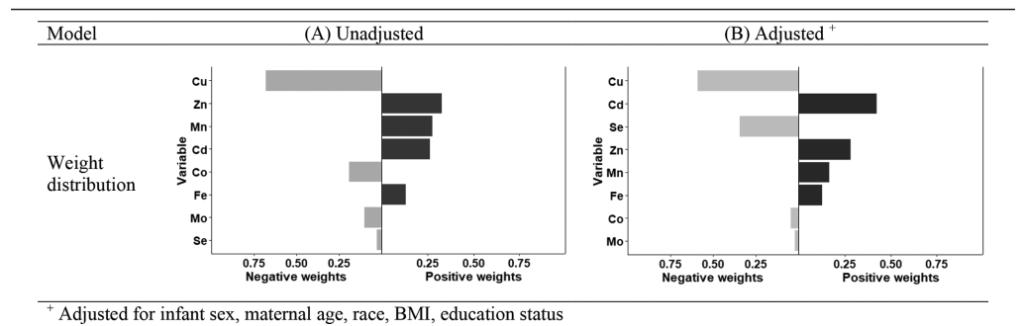
The Rhode Island study used something called the quantile g-computation approach to understand the association between metal mixture and NNNS profile 5 versus the other four profiles. The model assumes the linearity of simultaneously occurring metals some of which positively and some negatively affect the NNNS scores. Each exposure was given a positive or negative weight which add up to be 1. Cu showed the largest negative weight among the metals followed by selenium in the model adjusted for infant sex, age, and maternal race, BMI, and education status.
The Rhode Island study discussed the role of Cu in development as well as other studies rationalized the importance of copper in neurological development.
In Taiwan
A Taiwanese study pregnant women and their single birth children and correlated psychiatric functioning with heavy metals and phthalate esters in the urine. [5] Phthalate esters are a class of plasticizers with an increase in consumer exposure. The authors claimed that prenatal exposure to Cu, dibutyl phthalate, and di-2-ethylhexyl
phthalate was associated with child depressive problems and attention deficit/
hyperactivity problems at 4 years of age. [5]
confounding graphs
Smoking and drinking are sometimes considered confounding variables. People who smoke are more likely to drink alcohol. These authors used a directed acyclic graph (DAG, Figure S1) to illustrate the confounding variables that they needed to adjust for:
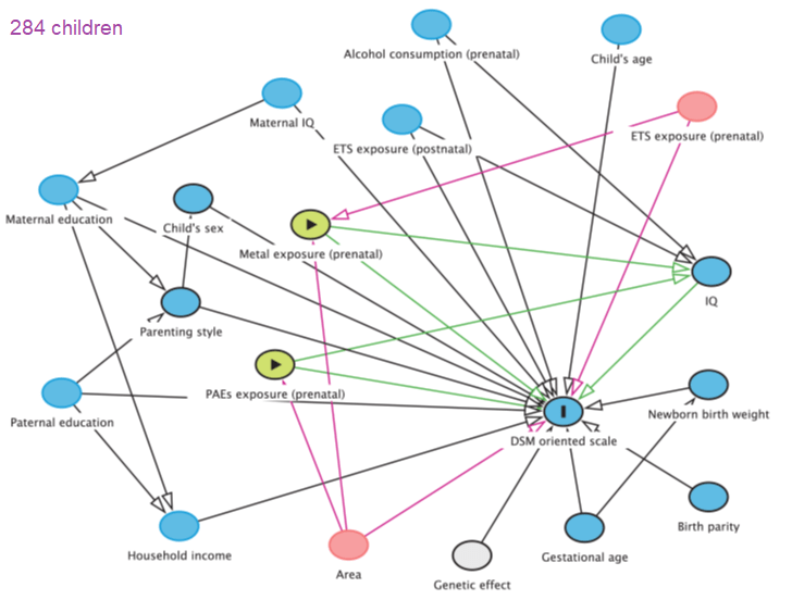
The blue oval with the solid bar is the outcome measure: The green ovals with the arrows are the two environmental factors hypothesized to lead to mental health problems as measured by the Diagnostic and Statistical Manual of Mental Disorders, Fifth Edition (DSM-5) All of the other ovals are confounding variables.
- prenatal environmental tobacco smoke ETS exposure (yes or no) and geographic area (central, southern, or eastern) for metals analysis, with adjusted geographic area for the analysis of PAEs.
- Model 1 adjusted for child’s age and sex (boy or girl), maternal education level (≤12 years, 12–16 years, and > 16 years), birth parity (1, 2, or ≥3), gestational age, prenatal ETS exposure, and geographic area
- Model 2 included the child’s IQ as measured by the Wechsler Preschool Primary Scale of Intelligence Fourth Edition
Spearman correlation coefficients
The Spearman correlation coefficient is a way of correlating the relationship between two variables, the closer the R value is to1.0 (direct) or to -1 (inverse) the stronger the correlation. This coefficient assumes the data are monotonic. An example is given of non-monotonic data.
The complexity of the situation is further illustrated in supplemental figure 2 which illustrates a rather sobering reality by presenting a table of Spearman correlation coefficients. All of the squares are green and blue indicating a positive correlation. The asterisk indicate the level of confidence. In simple lay terms * indicates that we are 95% confident that the observed correlation is not due to random chance, ** 99% confident.
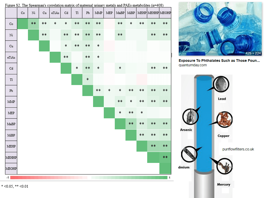
Of course a mother peeing one phthalate metabolite is likely to be peeing others. If a mother lives in a region in which she knows the water is contaminated with heavy metals, she may be more likely to drink bottled water and become exposed to phthalates as well. According to this table, there is a positive correlation between the concentration of lead (Pb) in urine with the concentration of copper ( Cu), nickel (Ni), cadmium, (Cd) cobalt ( Co ), and total inorganic arsenic (eTiAs).
phthalates and metals in combo

β (SE) may refer to the median 50% value of a quartile with SE being the standard error.
In statistics, p and q values are related entities. P values are the fraction of false positives whereas q values are the rate of false discovery. “To put this another way, p-values tell you the percentage of false positives to expect and take into account the number of tests being run. For example, if you run 1600 tests, you would expect to see about 80 false positives. The q-value doesn’t take into account all the tests; they only take into account the tests that are below a threshold that you choose (i.e. tests reporting a q-value of 5% or less).”
An odds ratio (OR) is the ratio of the odds of two events occurring simultaneously.
Spearman correlations and hypothetical graphs
Note that the graphs on the left are not real data from the study. They are being presented to give the lay reader an idea of what the real data might look like based on the R values.
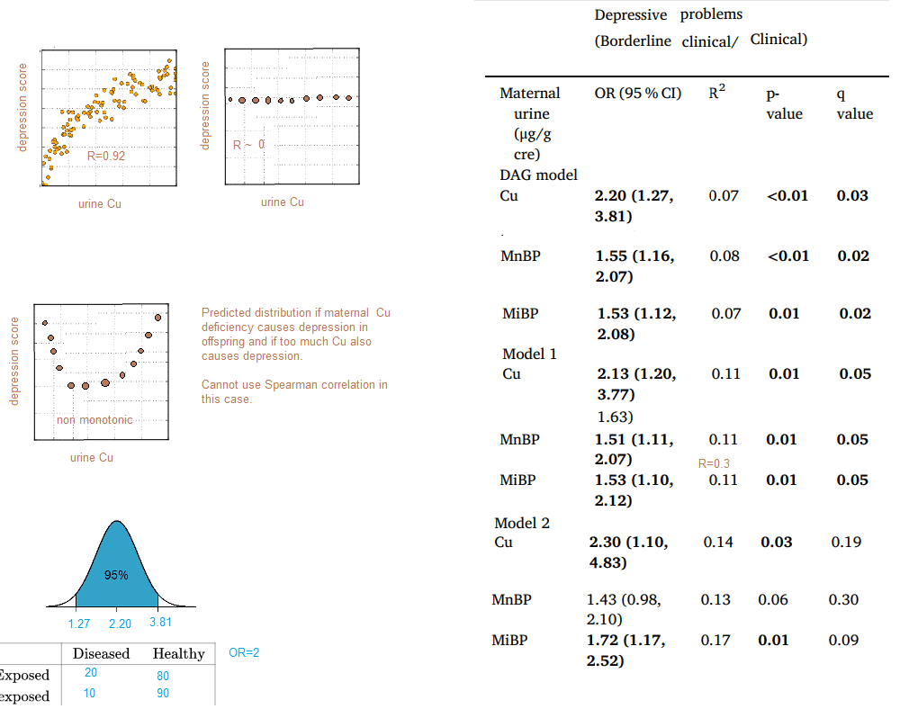
One of the things that makes this publication is that the authors assume that the reader is an expert in statistics and at the same time they don’t actually who their Spearman correlation data graphically. A search of the PDF file of the manuscript for “monotonic” did not reveal whether or not this assumption was met. Reporting R2 rather than R is the topic of considerable Internet debate. The bottom line is that the data were not known. The authors certainly seem to have a firm understanding of statistics. It would just be nice to see the data because too little copper is known to cause problems.
Other sites on prenatal copper
- Being the Parent brings up some recommended daily allowance and a list of copper rich foods.
- The Baby Center has similar information for expecting mothers and also advises a mother to talk to her doctor about the best supplement should her diet not contain enough copper.
- This Mayo Clinic site turned up on an Internet search but didn’t say anything bout copper supplements during pregnancy.
- What to Expect also gives the RDA for copper and a list of copper rich foods.
- MindBodyGreen turned up in an Internet search of prenatal copper for unknown reasons. These authors stated that it is possible to get to much zinc but say nothing about too much zinc leading to copper deficiency. [1]
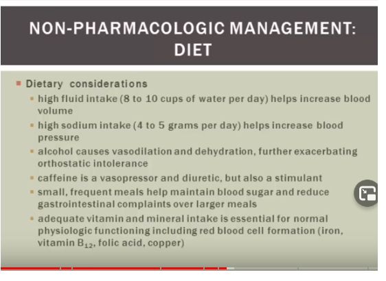
Talk to your healthcare provider
“Talk to your healthcare provider.” Is the obvious answer when sources of contaminants and getting proper amounts of trace elements seems to be overwhelming. Both the Rhode Island [4] and Taiwan [5] are public access for your healthcare provider to review with you.

Things are crazy complicated that seem to become only more complicated when statistical analyses are used. Surely there is a common sense approach going back to days in which are soils were not copper depleted and heavy metal and plastic contamination were unknown.
References
- Uriu-Adams JY, Scherr RE, Lanoue L, Keen CL. Influence of copper on early development: prenatal and postnatal considerations. Biofactors. 2010 Mar-Apr;36(2):136-52.
- Zimmerman AW, Matthieu JM, Quarles RH, Brady RO, Hsu JM. Hypomyelination in copper-deficient rats. Prenatal and postnatal copper replacement. Arch Neurol. 1976 Feb;33(2):111-9.
- Zirngibl M, Assinck P, Sizov A, Caprariello AV, Plemel JR. Oligodendrocyte death and myelin loss in the cuprizone model: an updated overview of the intrinsic and extrinsic causes of cuprizone demyelination. Mol Neurodegener. 2022 May 7;17(1):34. PMC free article
- Tung PW, Burt A, Karagas M, Jackson BP, Punshon T, Lester B, Marsit CJ. Prenatal exposure to metal mixtures and newborn neurobehavior in the Rhode Island Child Health Study. Environ Epidemiol. 2022 Jan 28;6(1):e194. PMC free article
- Tsai TL, Hsieh CJ, Wu MT, Chen ML, Kuo PH, Wang SL. Co-exposure to toxic metals and phthalates in pregnant women and their children’s mental health problems aged four years – Taiwan Maternal and Infant Cohort Study (TMICS). Environ Int. 2023 Feb 4;173:107804 free article