
We tend to accept that our cancer risks increase as we age. In introductory remarks on an exciting new landmark paper Li and coauthors were commenting on the fate of poor CD8+ T cells in the center of tumor micro environment (TME). CD8+ cytotoxic T cells are antigen specific recognizing antigens from tumors, virus infected cells, and normal cell. CD8+ is a protein that binds to the constant chain of the MHC1 antigen presenting surface protein. Recognizing normal cells is considered an act of autoimmunity. When exposed to infected/dysfunctional somatic cells, CD8+ T cells release the cytotoxins perforin, granzymes, and granulysin, enzymes that activate the caspase protease cascade eventually leading to programmed cell death. Human Cell Bio. Not only are the T cells constantly exposed to antigen that sets them off, but they are also competing with the tumor for oxygen. [1] Naturally reactive oxygen species are part of the TME. [2] According to Healthline, age is not one of the risk factors for autoimmune diseases. In autoimmunity CD8+ T cells are also constantly exposed to self antigen. [3] It is desirable that they become exhausted and less self- reactive. Both of these featured studies used T cells from mice that are allergic to an egg protein ovalbumin. These dual roles of CD8+ T cells was addressed in a review by Collier and coauthors. [4]. This post will not get into the PD1/PDL1 checkpoint inhibitors that have been discussed elsewhere on this site. We have covered the inner-relationship between NADH/NADPH (derived from niacin) and reduced thiols. This post examines the possibility that cuprous niacin could find the happy middle ground between T cell exhaustion in cancer and viral infections and autoimmunity, both of these may be issues as we age. Perhaps age is not an autoimmune disease risk factor, but surely contributes to the symptoms.
- Tales from the tumor micro environment [2]
- Nicotinamide slowing down CD8+OT-I T cells? [3]
- Concluding thoughts
- References
Tales from the tumor micro environment [2]
Polyclonal CD8+ T cells, T cells were isolated from the spleen and inguinal lymph nodes of C57BL/6 mice. T cells were also isolated from OT-I mice. These mice contain transgenic inserts for mouse Tcra-V2 and Tcrb-V5 genes. The transgenic T cell receptor was designed to recognize ovalbumin peptide residues 257-264 (OVA257-264) in the context of H2Kb (CD8 co-receptor interaction with MHC class I). The take home is that chronically stimulated T cells, by tumor antigens or ovalbumin, leads to impaired mitochondrial function and more reactive oxygen species generation. Both copper and niacin have the potential to improve this situation.

This results in MHC class I-restricted, ovalbumin specific, CD8+ T cells (OT-I cells). That is, the CD8 T cells of this mouse primarily recognize OVA257-264 when presented by the MHC I molecule. Cells were cultured in the presence of 1 μM of SIINFEKL peptide, a peptide from ovalbumin. The melanoma cells supplied the MHC class I to present the transgenic T cells with their ovalbumin peptide. Vardhana used B16-F10, B16-OVA, EL4, and A20 meleanoma cell lines. An interesting nuance is that they added 50 μM of the reducing agent β-mercaptoethanol (β-ME).
Figure 1, an increase in glycolysis
In the melanoma cells, 1 ng/mL IFN-γ was to induce MHC-I expression. IL-2, 10 ng/mL, was also added with (chronic) or without (acute) 1 μM SIINFEKL. T cells were passaged into fresh co-cultures every 48 h until eight days following initial stimulation with the peptide.
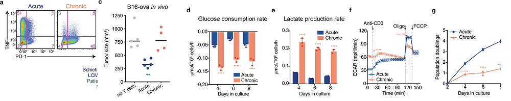
- In Figure 1a we see the same story: The more PD-1 on the cell surface, the less TNF in vesicles. It is the chronically stimulated cells that have the most PD-1. Skipping some flow cytometry data….
- Panel d, The chronically stimulated T cells consume much more glucose than the T cells only stimulated once with the ovalbumin peptide. This glucose could be for glycolysis only or glycolysis plus OxPhos.
- Panel e shows an increase in lactate production indicating that glucose consumption is probably not going towards OxPhos.
- Panel f demonstrates that the extracellular acidification rate (ECAR) is increased when both the chronic and the acute stimulated T cells are stimulated with CD3 antibodies. The acute stimulation cells acidify more in the presence of electron transport chain blockers. And finally,
- panel g, the acute stimulation T cells divide almost 4x as fast as those chronically stimulated with the ovalbumin peptide .
Fig 2 less O2 reduction and ATP production with chronic stimulation
Vardhala also used radioactive glucose to measure TCA cycle activity in these T cells (panels d-f not shown).. Some of the nucleotide ratios are not being shown in this post in effort to keep the narrative simple. It is just impossible to do this public access publication justice in a single post.
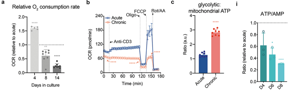
They saw a decrease in all intermediates in the TCA cycle. [2] Triphosphate and monophosphate nucleotide ratios were also reduced in the chronic stimulated T cells compared to the one time only acute stimulated cells. [2] Panel i from the chronic model documents that he problem gets worse with time.
Figure 3 mitochondria inhibition and T cell proliferation (not shown)
Cu(I)NA2 is predicted to supply Cu/Zn SOD with the necessary cofactor to mitigate the ROS generation as well as keep the flow of electrons through through the electron transport chain. Both of these should increase the ATP/AMP ratio as well as decrease the NADH/NAD+ ratios.
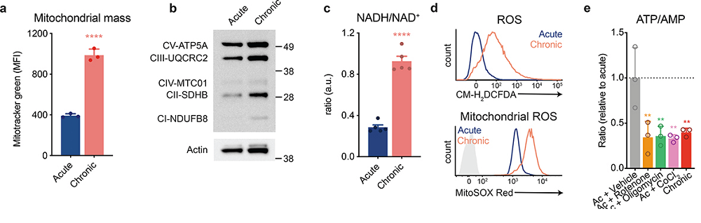
- Panel a Mitotracker green is simply a fluorescent indicator there to measure mitochondria size irrespective of activity. These mitochondria might be undergoing a vain attempt to expand and keep up with demand.
- panel b This is a Western blot. The black bands are proteins. There does not appear to b a huge difference in protein amounts of various complex proteins.
- panel c This is something we might want to copy. A smaller NADH/NAD+ ratio in the acute T cells that were not over exposed to peptide suggest better functioning mitochondria.
- Panel d examines the total and mitochondrial reactive oxygen species upon stimulation. The gray shaded trace is the unstimulated reference.
- Panel e Various inhibitors of electron transport chain complexes were used to mimic he effect of chronic stimulation: complex I (rotenone), complex V (oligomycin),Fe-S cluster containing complexes, cobalt chloride (CoCl2),
reactive oxygen species and reduced thiol supplementation
The rest of the Vardhana publication addressed the issue that the real root of reduced T cell proliferation was due to ROS generation rather than impaired mitochondria activity.
Nicotinamide slowing down CD8+OT-I T cells? [3]
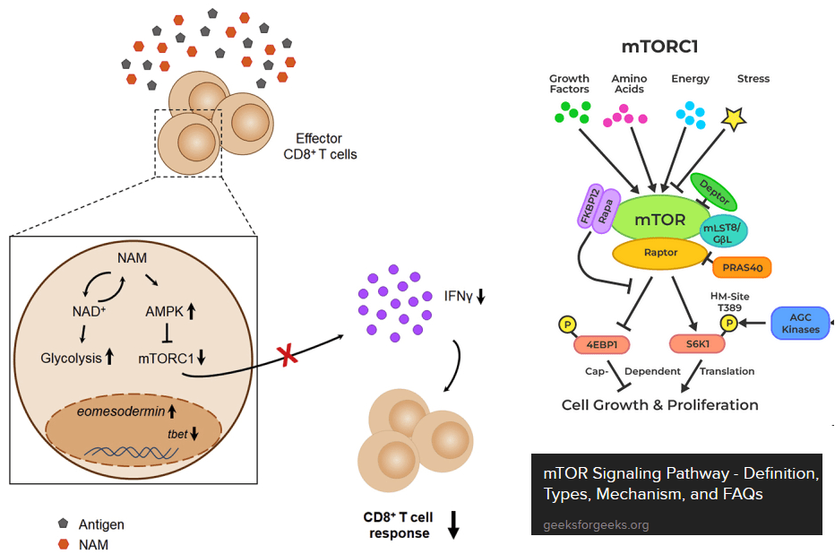
The next paper tests the hypothesis that nicotinamide can shut down over responsive CD8+ T effector cells. [3] Just as a warning, this is the exact opposite of T cell exhaustion. The paper makes use of the OT-I mice genetically engineered to think that the ovalbumin egg protein is a virus. These authors introduced us to the mammalian target of Rapamycin, or mTOR. mTOR integrates all sorts of signals from everywhere and gives the cell permission to start translating mRNAs into proteins. Ribosomal S6 kinase is a target of mTOR. This kinase promotes translation of mRNAs. Note that increases in the ratios of ADP to ATP tend to turn mTOR off and sufficient nutrients tend to turn it on. These authors used the Sea Horse to measure extracellular acidification rate, i.e. glycolysis without the TCA cycle and electron transport chain. [3]
Figures 1 and 2 Turning on cytokine production, or not…
Naïve CD3+, CD4+, CD8+ T cells from spleen and lymph nodes from C57BL/6J mice were purified and differentiated using a commercial kit with added 30 mM or 10 mM NAM or the solution used to dissolve the NAM. Final activation was with CD3/CD28 or PMA + ionomicyn (PMAi), a combo used to stimulate T cell activation, proliferation, and cytokine production. The authors saw differences in cytokine production, but by their own admission, used an surrealistically high concentration of NAM in their cell culture experiments. [3] Not getting into the specifics, the reason has to do with mTOR.
Figure 3 Some Sea Horse experiments
Let us continue with the Agliano [3] publication, which also used T cells from OT-I mice. Recall that these cells were engineered to react with the ovalbumin peptide provided that it is presented via MHC1 surface proteins.
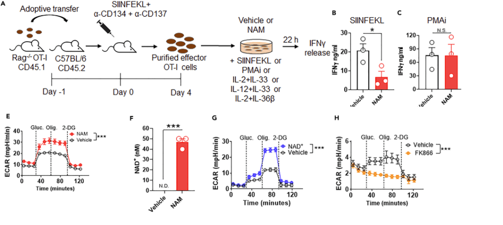
- Panel A the general protocol. They are activating the T cells with the peptide, phorbol ester, or a bunch of cytokines.
- Panels Band C Nicotinamide only slows down production of INFγ in response to the peptide, but not the phorbol ester plus ionomycin Ca2+ ionophore. PMA activates protein kinase C. ionomycin pokes Ca2+ permeable holes in cell membranes. This is sort of a double whammy way of activating T cells. Panel D examined response to interleukins.
- Panel E. Here Angliano and coauthors are comparing 10 mM nicotinamide (NAM) and the solution (vehicle) used to dissolve NAM. When glucose (gluc) is added the cell culture medium starts acidifying. Adding the ATP synthase inhibitor oligomysin (Olig.) does nothing. At 122 g per mole, an average human male with 5 liters of blood would have to consume about 6.1 g of nicotinamide to theoretically achieve this concentration.
- Panel F The large extracellular concentrations of nicotinamide are increasing the intracellular concentrations of NAD+.
- Panel G used only 1 mM NAD+. Consuming 0.6 g of NAD+ probably won’t happen either.
- Panel H, FK866 is a highly specific noncompetitive inhibitor of nicotinamide phospho ribosyltransferase . The authors presented another narrative suggesting that the large doses of niotininamide were slowing the translation of cytokine transcripts via phospho-AMP kinase pathways involving mTOR. [3]
Figure 4 demonstrates that NAM down regulates mTORC1 independently of NAD+. [3] Figures 5 and 6 looked at the effect of NAM on human T effector cells. This post is not going to get into this part of the study.
Concluding thoughts
The Vardhana paper made the cause for CD8+ T cell exhaustion being tied to reactive oxygen species and proposed use of reduced thiol supplements like N-acetyl cysteine. [2] We’d like to point out that (1) copper is a cofactor for Cu/Zn superoxide dismutase and (2) niacin is a precursor for NADH/NADPH. NADPH, in particular, plays a role in keeping thiols reduced. The Agliano paper made the case niacin’s close relative nicotinamide, being a good treatment autoimmune diseases when CD8+ T cells become cytotoxic against self, i.e. autoimmunity. [3] This site is in not way making any claims to treat disease. That cuprous niacin could treat two opposite ends of immune dysregulation would be an audacious claim and perhaps a worthy hypothesis to test!
References
- Li W, Cheng H, Li G, Zhang L. Mitochondrial Damage and the Road to Exhaustion. Cell Metab. 2020 Dec 1;32(6):905-907. free article
- Vardhana, S. A., Hwee, M. A., Berisa, M., Wells, D. K., Yost, K. E., King, B., Smith, M., Herrera, P. S., Chang, H. Y., Satpathy, A. T., van den Brink, M., Cross, J. R., & Thompson, C. B. (2020). Impaired mitochondrial oxidative phosphorylation limits the self-renewal of T cells exposed to persistent antigen. Nature immunology, 21(9), 1022–1033. PMC free article
- Agliano, Federica et al. “Nicotinamide breaks effector CD8 T cell responses by targeting mTOR signaling.†iScience vol. 25,3 103932. 15 Feb. 2022, free article
- Collier JL, Weiss SA, Pauken KE, Sen DR, Sharpe AH. Not-so-opposite ends of the spectrum: CD8+ T cell dysfunction across chronic infection, cancer and autoimmunity. Nat Immunol. 2021 Jul;22(7):809-819. PMC free article
Leave a Reply