This post is refuting an attack on Dr George Brewer who joined us in proclaiming the need for CopperOne. Dr Brewer claimed that small amounts of Cu2+ in our drinking water may contribute to Alzheimer’s Disease. This post asking if copper is safe for seniors critically examines the claims of Dr George Brewer.
The featured positron emission tomatography (PET) study loaded the blood mice with 64Cu2+ acetate a cell permeable Cu+ chelaate and monitored its distribution over a 24 hour period. They loaded some TASTPM mice engineered with gene mutations associated with early onset Alzheimer’s Disease. [1] Towards the end, the special TASTPM mouse Alzheimer’s Disease model and copper’s role in amyloids will be discussed.
Argument against Dr Brewer
Point 1. This post has nothing to say about the cost. This post will review 2022 literature on the link between copper II Cu2+ and Alzheimer’s Disease so that the reader will understand the cost more.
Point 2. Their hypothesis:
At the Mitosynergy product page above is a link to what they claim is the science behind their product:
by George J Brewer
A similar article by George Brewer can be found here:
https://www.scientia.global/professor-george-brewer-how-to-avoid-alzheimers-disease/
“I refuted at least two of these “anti copper†studies in my book. First, 0.12 ppm copper in water is the same as 0.12 mg/Liter. It’s not copper to blame, it’s clearly copper deficiency. It is not nearly enough copper to be toxic.
In the second study, it’s 3 mg of copper. Paired with known neurotoxic bad fats, and a diet very high in those bad fats. Again, not enough copper to be toxic, but the copper does help our bodies turn fats into the myelin sheath around the nerves, which is why, even in our Facebook group’s description, at https://www.facebook.com/groups/thecopperrevolution”
Interesting points, Jason Hommel. Let’s explore where this Cu2+ was found to accumulate.
Prelude: ventricles and copper
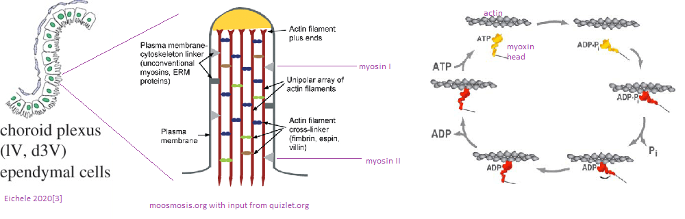
As we shall discuss, Cu2+ becomes deposited in the ventricles of the brains of normal middle aged and old mice. [1] Since at least 1988 we have known that the ciliated ependymal cells of the ventricles contain actin and myosin. [2] Skeletal muscle isoforms of actin and myosin are responsible for movement of those muscles. Likewise, these ciliated cells are absolutely required for moving cerebral spinal fluid about the brain. [3] Myosin hydrolysis of ATP is required for such movement. ATP is most efficiently produced by the electron transport chain of the the mitochondria. In Alzheimer’s disease, a decrease in expression of complex IV genes COX6A1, COX6C, COX11, SOD1, ATOX1, COX5B, COX6B1, and COX7B has been observed. [4] A decrease in transcripts for Cu/Zn super oxide dismutase 1 and copper chaperone Atox1 were also observed. [4]
If we do not have enough proper copper in our diets, we could hypothetically not have enough to ATP to fuel the intense ventricular circulation dynamics reviewed in reference [3]. In this model we are proposing, not enough of the good copper Cu+ can lead to accumulation of the bad Cu2+ before it has a chance to be reduced to the good Cu+. With that introductory prelude, let us examine a PET imaging study of Cu2+ acetate in a mouse model.
Brain imaging of 64Cu
1. Like Cu2+ from pipes and cell membrane permeable Cu+/2+
The goal was to directly inject two forms of Cu(II) into the blood: one in a GTSM chelate that can cross the blood brain barrier and one that might resemble a dietary supplement. [1]
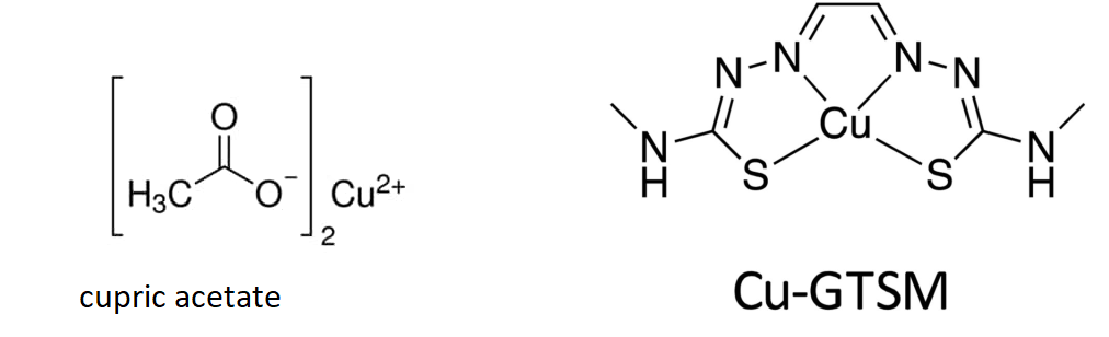
The authors are trying to load the mice with two types of copper. (1) copper pipes copper and (2) copper in the mostly proper oxidation state with a lipophilic carrier that can cross the blood brain barrier.
2. Cu2+ exit from blood
Both copper compounds were IV injected in the tail veins as opposed to feeding. Radioactivity was measured over the course of 60 minutes. The radioactivity in the blood was expressed in terms of the ratio of counts per minute (cpm) to the MBq injected. (n = 3 ) 1 Bq = 1 decay per second. Initial clearance of Cu-GTSM is very rapid with half life < 1 min, whereas Cu-acetate had a slower initial clearance half life of 2-3 min. [1]
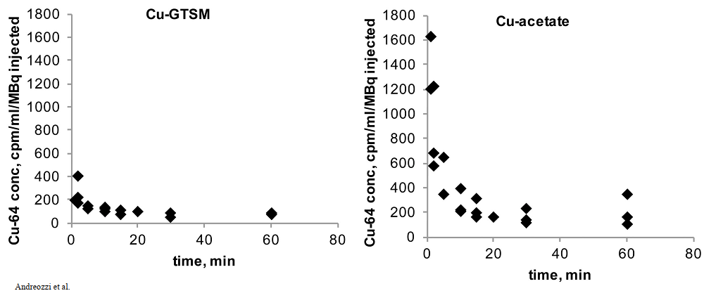
The cupric acetate seems to be higher in the blood. Was this due to slower deposition into disposition into tissues or quicker excretion in feces and urine for the GTSM-Cu? This is addressed in Figure 3 at 30 minutes. Note the increased signal from the intestines and kidney.
3. Improper copper gets peed out?
These are the wildtype C57BL/6J mice after injection with the two forms of 64Cu. Both forms are rapidly taken up by the liver, intestines, and kidneys. Is the presence in the liver and kidneys evidence of being excreted? The GTSM chelate is also taken up into the brain, heart, lungs, and adrenal glands. The color scale is linear, covering the range 0-1 %ID/mL (min) to 40 %ID/mL (max).
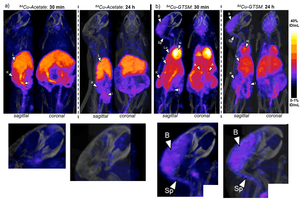
Some enlarged views of sagittal brain sections are shown. The brain (B) and spinal cord(Sp) rapidly accumulate 64Cu from the chelate.
The heat, lung, and brain are the biggest difference between the cupric acetate and GTSM, that tends towards the Cu(I) oxidation state. The brain seems to unload the accumulated Cu from 30 minutes to Why the difference between the brain and heart at 24 hours (p<0.001) at 24 hours is so important was not really elucidated. The 30 minute accumulation at 30 minutes is lost by 24 hours. Not so in the brain.
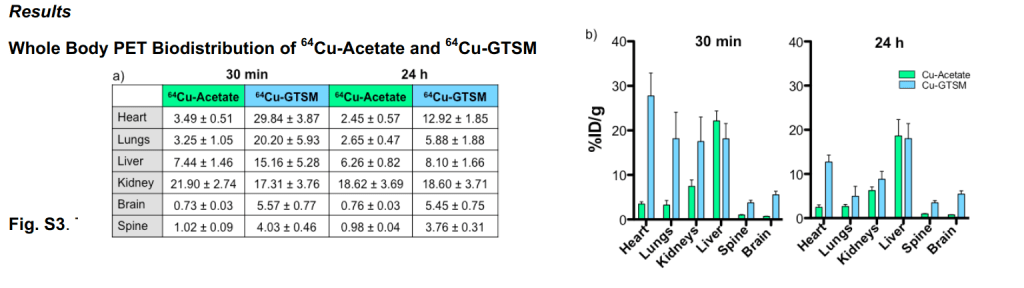
With the unnaturalGTSM Cu(I) chelate, the Cu peaks and then drops dramatically in most organs other than the brain, spine, and kidney over the course of 24 hours.
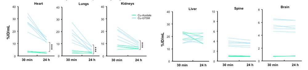
4. Copper getting stuck in the brain and spine
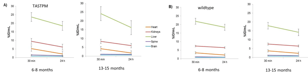
The supplemental data included individual mouse data from 30 minutes and 24 hours. These again are youngish 6-8 month old wild type mice.
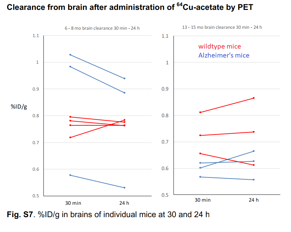
The authors took a closer look at Cu2+ acetate in 6-8 month old mice and some very old 13-15 month old mice. [1] While they only looked at three very old wild type mice, one individual had a slight increase in brain Cu over 24 hours. We can make no statistical conclusions from such a small group. This is sort of what Dr Brewer was saying that Cu2+ might just accumulate very slowly in small amounts as we age. Note also that there is greater population variation in the older versus younger group of wildtype mice.
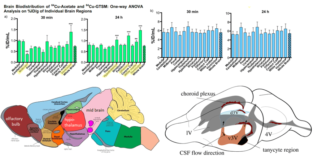
This table breaks down the 64Cu content by brain region
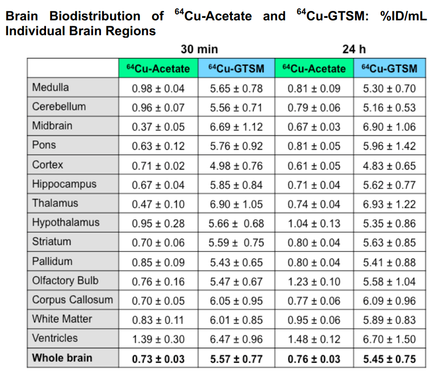
Eichelle (2019) [3] was the source of the rodent brain ventricle system. Note that the d3 ventricle and choroid plexis line up with regions in which Cu content is increased with Cu2+. The choroid plexis produces cerebral spinal fluid and serves as a barrier between the blood and the cerebral spinal fluid in the ventricles
And there was really not a great deal of difference in the Alzheimer’s Disease model
TASTPMouse
This Adreozzi study [1] also used what is known as a TASTPM mouse model of human Alzheimer’s Disease. [1] This particular mouse like carries two different sets of mutations linked to early onset Alzheimer’s Disease in humans.
- The “Swedish†mutation in the APP gene is two mutations K595A and M596L that increases cleavage by β-secretases
- An M146V substitution in the presenilin gene. According to alzforum.org, this mutation impaired γ-cleavage carboxypeptidase like activity but spared the endopeptidase ε-cleavage. This results in increased Aβ1-42 compared to Aβ1-42
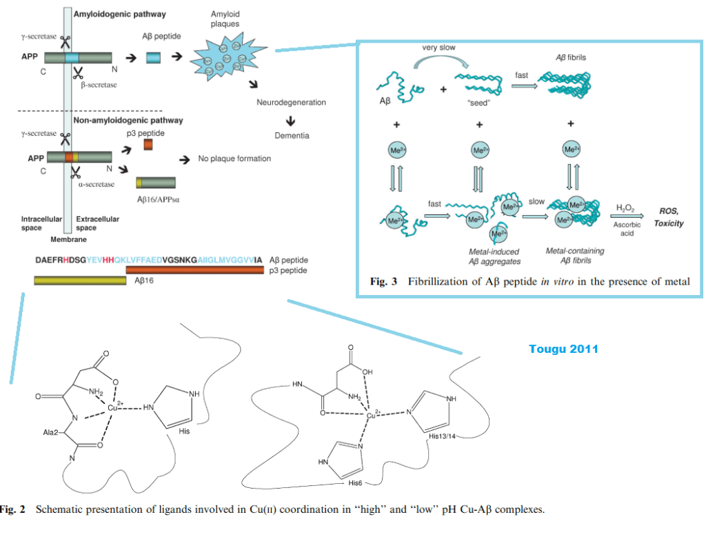
The Tougu (2011) kinetic study [2] offered some insight into the role of divalent metal ions in promoting the aggregation of the Aβ peptides and the histidines therein. Their figure 1 puts into context the role of secretases and the differences between proteolytic cleavage products. Figure 2 has the metal binding histidines in bold red letting us know that the proteases really make a difference. [2] Figure 3 shows the different pathways to aggregates that generate reactive oxygen species in the presence of ascorbate and the H2O2 that seems to be the byproduct of incomplete reduction of O2 by the mitochondria.
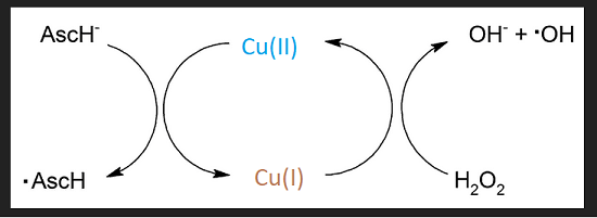
There have been other reports claiming Cu2+ detection by one means or another. [3-5] The Tougu Figure 3 reminds us that Cu2+ and Cu+ redox cycle with ascorbate and H2O2 to produce hydroxyl radicals.
In Alzheimer’s Disease, there is a decrease in the transcripts for many of the subunits of the copper cofactor mitochondrial complex IV. Transcripts for Cu/Zn SOD1 and the copper bound transcription factor and chaperone Atox1 are also decreased. These changes are accompanied by a decrease in Cu, a decrease in cytochrome C oxidase activity, and of course a decrease in ATP production. [6]
Going forward..
The GTSM mostly Cu+ is unnatural by nature of its cell permeability and therefore not of interest to us.
- Can we detect impaired mitochondrial function in the aging brain?
- Does Cu(I)NA2 improve CSF circulation within the brain?
- Is Cu2+ less likely to form aggregates withAβ if CSF is moving freely and not stagnating?
The Andreozzi study was performed over only 24 hours. What about dietary Cu2+ over the course of several decards?
References
- Andreozzi, E. M., Torres, J. B., Sunassee, K., Dunn, J., Walker-Samuel, S., Szanda, I., & Blower, P. J. (2017). Studies of copper trafficking in a mouse model of Alzheimer’s disease by positron emission tomography: comparison of 64Cu acetate and 64CuGTSM. PMC free article
- C, Ghandour MS, Paulin D, Assenmacher I, Tixier-Vidal A. Characterization of ependymal cells in hypothalamic and choroidal primary cultures. Neuroscience. 1988 Mar;24(3):993-1007
- Eichele, G., Bodenschatz, E., Ditte, Z., Günther, A. K., Kapoor, S., Wang, Y., & Westendorf, C. (2020). Cilia-driven flows in the brain third ventricle. Philosophical transactions of the Royal Society of London. Series B, Biological sciences, 375(1792), 20190154. PMC free article
- Myhre, O., Utkilen, H., Duale, N., Brunborg, G., & Hofer, T. (2013). Metal dyshomeostasis and inflammation in Alzheimer’s and Parkinson’s diseases: possible impact of environmental exposures. Oxidative medicine and cellular longevity, 2013, 726954. PMC free article
- Tõugu V, Tiiman A, Palumaa P. (2011)Interactions of Zn(II) and Cu(II) ions with Alzheimer’s amyloid-beta peptide. Metal ion binding, contribution to fibrillization and toxicity. Metallomics. 2011 Mar;3(3):250-61. free article
Leave a Reply