This post examines the rat experiments in this paper that seems to have made a CopperTwo version of CopperOne and is claiming that CopperTwo has skin healing properties. The first two posts in this series summarizes the production protocol and chemical characterization followed by some fibroblast and endothelial cell culture studies. The rat studies might have been meant to “bring home the gold.” Sadly, much of the histology has not been quantitated. The readers are left to decide for themselves if the changes are meaningful. The purpose of this post is to allow those with financial interest in CopperOne for wound treatment if these CopperTwo results are enough to warrant further pursuit.
Wang TL, Zhou ZF, Liu JF, Hou XD, Zhou Z, Dai YL, Hou ZY, Chen F, Zheng LP. Donut-like MOFs of copper/nicotinic acid and composite hydrogels with superior bioactivity for rh-bFGF delivering and skin wound healing. J Nanobiotechnology. 2021 Sep 9;19(1):275. PMC free article
Full‑thickness skin defect model and treatment
Female Sprague-Dawley rats were subjected to surgical incision after shaving hind skin of fur and other measured to minimize pain and infection. Two full thickness incision wounds (10 mm × 10 mm) were made on both sides of each rat. The defects were covered with GelMA 5% CuNA@GelMA and 5% CuNA-bFGF@GelMA and fixed with 2M medical tape. Wounds treated with only medical tape served as the negative control. Rats were euthanized on days 3, 7 and 14. The wound and normal
adjacent skin were excised and then fixed in 4% paraformaldehyde.
Wound closure measurement
The wounds were photographed with a digital camera after 3, 5, 7, 11 and 14 days and the wound area was measured by ImageJ software. The percentage of wound closure was defined as:
Wound closure(%) = (1 − P/I) × 100,
where I is the initial wound area and P is the wound area at a given time point. Each treatment group had three samples.
7. Histological evaluation
Most evaluations were performed using commercial kits and self-explanatory antibodies. . To identify elastic fibers, Weigert’s elastic staining was used to stain the sections. Histological analysis of wounds was performed on day 3, 7, and 14 to evaluate the impact of CuNA-bFGF@GelMA on wound healing. H&E and Masson’s trichrome staining was conducted after 3, 7 and 14 days after treatment. H&E stains nuclei purple, collagen shades of pink, and other cellular materials shades in between.
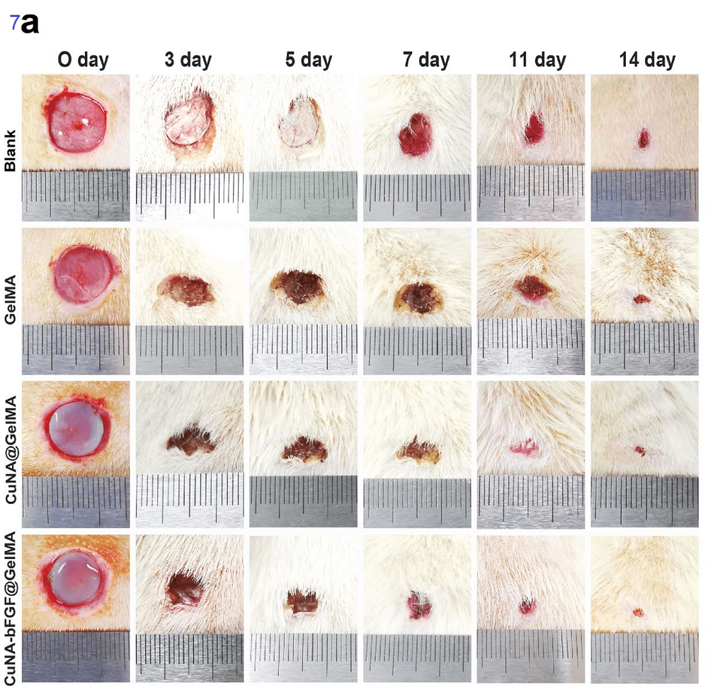
CuNA‑bFGF@GelMA on day 0, 3, 5, 7, 11 and 14.
- 7a All wounds shrank during each time period; however, the wounds treated with CuNA@GelMA and CuNA-bFGF@ GelMA shrank more. CuNA-bFGF proved to be particularly effective in promoting wound closure. The authors claimed a synergistic effect of copper and bFGF.

- 7b Data are expressed as mean±SD (n=3). (*p < 0.05, **<0.01, **<0.001 compared to the blank) The biggest improvement appears to be in the first three days.
- 7c Normal tissue stained with H&E. Note the blood vessels and regular epithelium.

7d, H&E staining reveals that CuNAbFGF@GelMA treatment results in skin closest to normal skin. The control and GelMA skin samples show basic epithelium and groups form the basic structure of epithelium and dermis but lack hair follicles, regular epithelium, new blood vessels, and the milder inflammation found in the CuNA and CuNAbFGF@GelMA treated skin responses. Especially the H&E staining of the group is closer to the normal tissue
8 Inflammation,epithelia, and blood vessels
This figure introduces two proteins of interest in wound healing. Interleukin 6, IL-6, is a pro inflammatory cytokine that may be produced by blood vessel smooth muscle cells. Ki-67 is a nuclear protein associated with cell proliferation. CD-31, aka pecam1, is in simple terms a marker of angiogenesis. In their cell culture experiments, these authors examined non muscle actin in endothelial cells and fibroblasts. α- Smooth muscle actin in this case is used as a marker of new blood vessel formation.
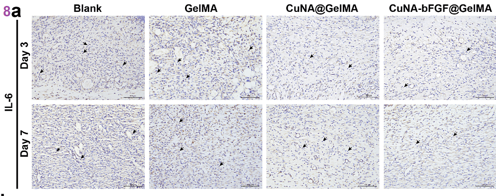
8a The blank and @GelMA show more IL-6 than the CuNA and NuNA&bFGF embedded in the @GelMA matrix. On day 3. In this case the small, brown, blobs seem to be the regions of positive IL-6 staining.
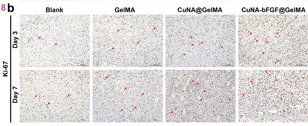
8b Ki-67 positive nuclei represent the re-epithelial-ization process. Figure 8b shows that a small number of Ki-67. CuNA-bFGF@GelMA group exhibits the highest level among all the groups.

8c CD31 (red) and α- smooth muscle actin (green) and nuclei (blue) While actin is a “house keeping†protein expressed in large amounts of most every tissue, the smooth muscle cells of blood vessels express a different isoform than fibroblasts and endothelial cells.
Readers are left to decide for themselves as to whether or not the changes in figure 8 are consequential because no numbers are given.
9. Collagen deposition
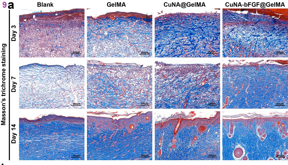
9a The authors were interested in a report that collagen formation is due to the massive proliferation of fibroblasts. Collagen deposition was examined in the wound regions using Masson’s trichrome staining which demonstrated much more collagen deposition in the CuNA-bFGF@GelMA treated skin. Masson’s trichrome stains as follows: red keratin and muscle fibers, blue or green collagen and bone, light red or pink cytoplasm, and dark brown to black cell nuclei.
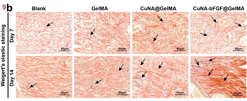
9b Weigert’s elastic staining was performed on day 7 and 14. The CuNA-bFGF@GelMA treatments
Leave a Reply