Author: BL

  • Aging, telomeres, hair

    We had thoughts, at one time, of measuring telomere length in cultured fibroblasts as a index of aging. Why not cultured hair follicles since so many customers report restoration of pigmentation in their hair when consuming BioCu1. Hair follicles are also more mitochondria reliant for their daily activities… unless the aging process is taking its toll. The more we thought about the proper controls, the harder and more expensive things seemed. Perhaps we are over thinking the story.

    1. Background Cu1 vs Cu2, don’t forget niacin
    2. Oxidative stress and telomere length
    3. Exposure to Cu1 and Cu2
    4. Aging studies: hair follicles in culture
      1. hair follicles are complicated organs
      2. Reactive oxygen species (ROS) not defined
    5. Follicles from hairs that have turned dark
    • Ctr1 describes the Ctr1 Cu(I) transport channel. Cu(II) does not get transported until it is reduced to Cu(I). We have always assumed that Cu(I)niacin is more bioavailable than Cu(II) sulfate.
    • Copper chaperones deliver Cu in the +1 oxidation state to their target enzymes. This is why we always thought, but never proved, that our copper is better.
    • Niacin in the Covid describes how we began to realize that Cu(I) and niacin likely work together.
    • Fatty Liver in Cows expands on the interplay between niacin and copper.

    von Zglinicki T. Oxidative stress shortens telomeres. Trends Biochem Sci. 2002; 27: 339-344 Sci-Hub free paper

    Some images were imported of the position of telomeres in a chromosome and examples of telomere shortening with cell divisions. Stem cells, red, keep a fairly constant telomere length. The moment a cell differentiates into a somatic cell (green) with a designated function, telomeres start to shorten. The Hayflick limit (HL) is the number of times a cell may divide before division stops. Crises (blue) may alter gene expression and increase telomere length. PD is the population doubling. (b) The downward green arrows in the von Zglinicki Fig 1 indicates that stress can shorten telomere length more rapidly than normal population doubling. (c) High stress and/or lack of good oxidative defenses, indicated by the width of the blue triangle, can send the telomere length into an almost vertical downward spiral. Good oxidative stress defense can send the telomere length response to

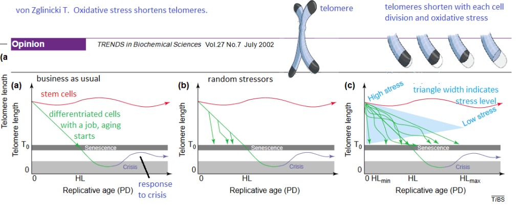
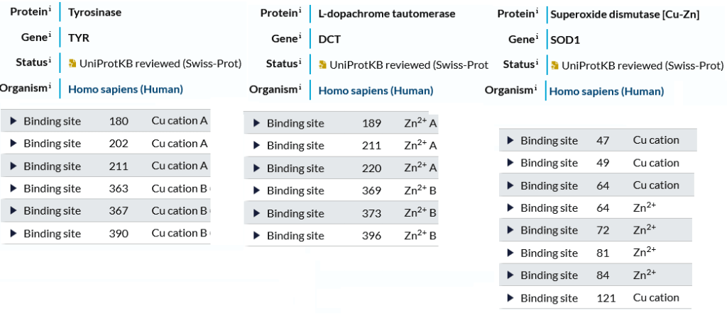
    Though this review was published over 20 years ago, it was very insightful and extremely useful for generating hypotheses. The Cu(I) of BioCu1 supplies Cu/Zn superoxide dismutase with a necessary cofactor. Niacin can be a precursor of NADH and NADPH, a cofactor for many anti-oxidant enzymes.

    Exposure to Cu1 and Cu2

    These are some thoughts on how we might design a cell culture experiment in fibroblasts or maybe even hair follicles in culture. Fibroblasts from individual patients and hair follicles could be studied.

    1. For starters we will need the culture medium without red dye that is used to indicate pH. We will add the same molar amount of Cu1 used in previous cell culture studies to the culture medium. We will take UV/Vis spectrophotometer scans every one minute for 15 minutes. We will calculate the time window in which we can keep the Cu1 in culture medium on the cells with less than 10% being oxidized to Cu2. (Cupric copper is blue).
    2. We could add both coppers to cultured follicles and/or fibroblasts for the designated time.
    3. After the incubation period, the copper containing media could be moved and the copper adhering to the outsides of the cells rinsed away.
    4. As another control for the Cu1 , we might want to consider the same molar amount of niacin without the copper. As a control for the cupric sulfate we might want to add a non copper sulfate.
    5. Do we have a way of measuring the copper that was taken up by the cells?
    6. The next step might be to measure ATP content.

    This is already starting to seem like a considerable amount to perform fibroblast or cultured hair follicle experiments. Others are using hair follicles in culture to study telomere aging.

    Stone RC, Aviv A, Paus R. Telomere Dynamics and Telomerase in the Biology of Hair Follicles and their Stem Cells as a Model for Aging Research. J Invest Dermatol. 2021 Apr;141(4S):1031-1040. free article

    • This review proposed that telomere length dynamics play an important role in the biology of the hair follicle (HF),
    • HF are mini organs that show an intriguing aging pattern in humans.
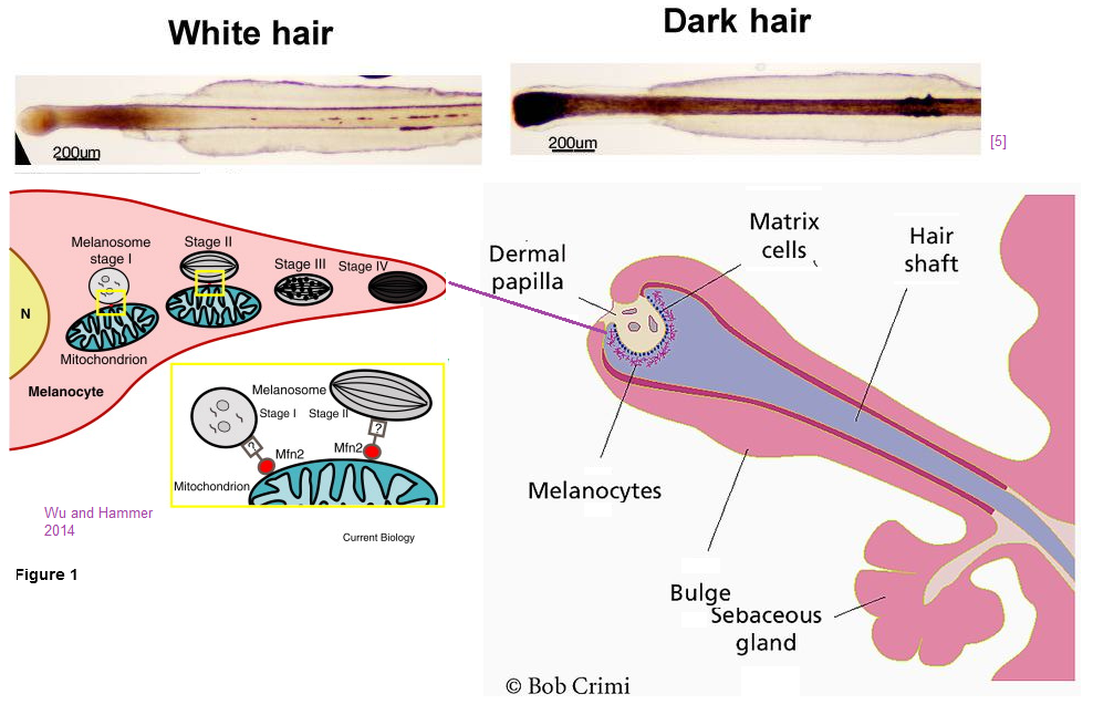
    • The pigment producing unit ages quickly but epithelial stem cells (ESC) are more aging resistant. Telomerase deficient mice with short telomeres display an aging phenotype of hair graying and hair loss that is attributed to impaired HF ESC mobilization.
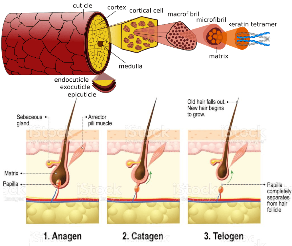
    Figure 1 Aging resistance in the cycling human HF. Anti oxidative capacity of the human HF is attributed to ROS scavenging molecules (e.g., catalase, melatonin), oxidative damage response controls (e.g., NRF2), synthesis of neuro hormones influencing mitochondrial function (TRH/TSH), and various DNA repair mechanisms. Similarly, the roles for telomerase and IGF-1 are proposed in the context of TL and TA in the HF. HF, hair follicle; NRF2, nuclear erythroid factor 2- related factor 2; TA, telomerase activity; TL, telomere length;

    The lay reader, and even trained biologists, would have to do so much background reading to properly understand what is being said in this review. This review does not discuss the anti-oxidant enzyme Cu/Zn super oxide dismutase simply because no one can think of everything at once.

    Figure 2 Proposed roles of TL in human HF biology and pathology.
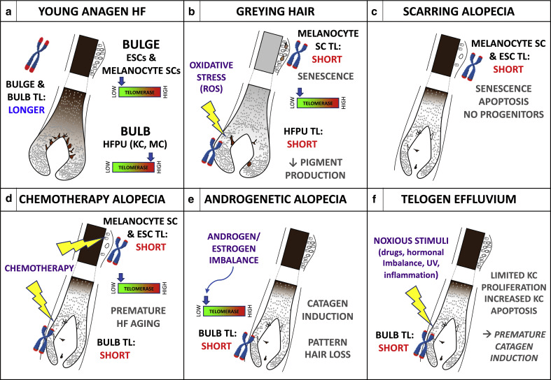
    (a) Young anagen HF with longer telomeres and low TA in the bulge region containing ESC, melanocyte SC and longer telomeres but high TA in the proliferative bulb region containing KC and MCs of the HFPU. (b) In graying hair, bulge melanocyte SCs with short telomeres senesce and limit new HFPU formation, whereas the ROS-sensitive aging HFPU accumulates endogenous and exogenous oxidative damage that shortens telomeres and impairs pigment production; induction of TA might prevent age-associated graying.(c) In cicatricial (scarring) alopecia, critically short telomeres in bulb SCs trigger senescence and apoptosis; with no progenitors and transient-amplifying cells, HF cycling and hair growth are irreversibly halted.
    (d) Chemotherapy (e.g., taxanes) damages telomeres and/or low TA HF SCs and also shortens telomeres of the stress-sensitive HFPU. Pretreatment with TA inducers might mitigate chemotherapy-induced HF aging and/or graying and hair loss. (e) Low HF aromatase activity may lower local estrogen and/or progesterone levels; loss of transcriptional stimulation of hTERT production leads to shortened telomeres and premature HF entry into catagen, contributing to AGA. (f) Noxious stimuli shorten hair matrix KC telomeres, limiting cycling of transient-amplifying cells and increasing apoptosis, leading to anagen termination and/or catagen induction and telogen effluvium. AGA, androgenetic alopecia; ESC, epithelial stem cell; HF, hair follicle; HFPU, hair follicle pigmentary unit; KC, keratinocyte; MC, melanocyte; SC, stem cell; TA, telomerase activity; TL, telomere length.

    This is all very interesting, but we’ve got to

    We have so many anecdotal stories of customers who have had their original hair color return after taking BioCu1. Why not just analyze the follicles from such hairs that have turned dark and compare them with hair follicles from the same customer before BioCu1.

  • Hair Growth

    For an overview of gray hair and a few works on the mitochondria requirement to grow hair, visit the gray hair post. The starting point of this post is Women’s World 8 essential oils for hair growth. The strategy is to do a PubMed search on each and then figure out which phytochemicals are in each and what their receptors are.

    Rosemary oil

    It took six months to see a difference in androgen associated hair loss. [1]

    1. Panahi Y, Taghizadeh M, Marzony ET, Sahebkar A. Rosemary oil vs minoxidil 2% for the treatment of androgenetic alopecia: a randomized comparative trial. Skinmed. 2015 Jan-Feb;13(1):15-21. PMID: 25842469. PubMed

    pumpkin seed oil

    In a 3 month clinical trial PSO showed promise in treating female pattern hair loss. In this 24 week clinical trial with men with androgen alopecia Mean hair count increases of 40% were observed in PSO-treated men at 24 weeks, whereas increases of 10% were observed in placebo-treated men (P < 0.001). Adverse effects were not different in the two groups.

    The phytochemical in question for pumpkin seed oil appears to be a phyto steroid testosterone mimetic [3]
    1. Ibrahim IM, Hasan MS, Elsabaa KI, Elsaie ML. Pumpkin seed oil vs. minoxidil 5% topical foam for the treatment of female pattern hair loss: A randomized comparative trial. J Cosmet Dermatol. 2021 Sep;20(9):2867-2873 PubMed
    2. Cho YH, Lee SY, Jeong DW, Choi EJ, Kim YJ, Lee JG, Yi YH, Cha HS. Effect of pumpkin seed oil on hair growth in men with androgenetic alopecia: a randomized, double-blind, placebo-controlled trial. Evid Based Complement Alternat Med. 2014;2014:549721. PMC free article
    3. Teeranachaideekul V, Parichatikanond W, Junyaprasert VB, Morakul B. Pumpkin Seed Oil-Loaded Niosomes for Topical Application: 5α-Reductase Inhibitory, Anti-Inflammatory, and In Vivo Anti-Hair Loss Effects. Pharmaceuticals (Basel). 2022 Jul 27;15(8):930. PMC free article

    peppermint oil

    Only one abstract was found on PubMed which focused on a type of hair loss specific to African American women which stated that there was not great evidence that this substance was effective.

    Ezekwe N, King M, Hollinger JC. The Use of Natural Ingredients in the Treatment of Alopecias with an Emphasis on Central Centrifugal Cicatricial Alopecia: A Systematic Review. J Clin Aesthet Dermatol. 2020 Aug;13(8):23-27. Epub 2020 Aug 1. PubMed

    lavender oil

    Also see the thyme oil section for the Turkish combination study.

    clary sage oil

    I could find nothing on PubMed concerning clary sage oil and hair loss. Titles indicate it is calming.

    cedarwood oil

    Also see the thyme oil section for the Turkish combination study.

    tea tree oil

    A study out of Saudi Arabia tested the hypothesis that adding tea tree oil would improve hair growth in male androgenic alopecia. These authors used a multimodal microemulsion of minoxidil (a dihydrotestosterone antagonist), diclofenac (a nonsteroidal anti-inflammatory agent), and tea tree oil which they viewed as an anti-infective agent. This publication reports stability factors that might be of interest to Mito. The good news is that significant hair growth was recorded after 32 weeks of use compared to the placebo and traditional medication.

    Farouk Sakr F, Gado A, Mohammed H, Ismail AAN. Preparation and evaluation of a multimodal minoxidil microemulsion versus minoxidil alone in the treatment of androgenic alopecia of mixed etiology: a pilot study. Drug Des Devel Ther. 2013;7:413. [PMC free article]

    thyme oil

    The Ezekwe review is the only thing that showed up on PubMed {1] This is not to say the research has not been performed. The Ezekwe review lead to a Turkish study of a combination of essential oils that are to be rubbed into the scalp. [2] The active group in this clinical trial received a compound available in Turkey: Revigen® Areata solution (Mikro-Gen Co, Istanbul, Turkey) which has essential oils, Thyme vulgaris, Lavandula agustifolia, Rosmainus officialis and Cedrus atlantica and mixed in carrier oil which was a combination of jojoba and grape seed oils.

    1. Ezekwe N, King M, Hollinger JC. The Use of Natural Ingredients in the Treatment of Alopecias with an Emphasis on Central Centrifugal Cicatricial Alopecia: A Systematic Review. J Clin Aesthet Dermatol. 2020 Aug;13(8):23-27. Epub 2020 Aug 1. PMC free article
    2. Özmen I, Çalişkan E, Arca E et al. Efficacy of aromatherapy in the treatment of localized alopecia areata: a double-blind placebo controlled study. Gulhane Med J. 2015;57:3. free article

    If the particular source of thyme oil contains the terpene thymol it might be effective as a skin permeability enhancer to get active ingredients past the stratum corneum.

    Holistic approach: biotin, niacin,more

    A very recent review mentioned popular use of biotin in products for hair regrowth but seemed to be much more optimistic about niacin. [1] The corresponding author of this paper is Raja Sivamani MD, who usually publishes with four or five institutes as his home base. These include a medical school and several research centers. Raja Sivamani has been part of numerous clinical trials.

    College of Medicine, California Northstate University, 9700 W Taron Dr, Elk Grove, CA 957
    Integrative Skin Science and Research, 1495 River Park Drive, Sacramento, CA
    Pacific Skin Institute, 1495 River Park Dr Suite 200, Sacramento, CA
    Department of Dermatology, University of California-Davis, Sacramento, CA

    Here is what the review [1] has to say about B vitamins:

    B Vitamins

    B vitamins, including niacin (vitamin B3), biotin (vitamin B7), and folic acid (vitamin B9) have been implicated in hair loss. For example, in addition to the well-documented pellagra characteristic of niacin deficiency, alopecia is an additional common clinical finding associated with deficient niacin [43]. However, no studies have directly assessed niacin levels among patients presenting solely with hair loss, and studies have found no significant difference in folate levels between alopecia patients and control subjects [57,58].

    “Biotin, a cofactor for carboxylation enzymes with dietary sources including protein, has been more extensively assessed for effects on hair parameters, and it is included in a variety of supplements or serums intended for hair health [44]. Genetic biotin deficiency is associated with severe dermatitis and alopecia (infantile) and sparse or absent scalp, eyebrows, and eyelash hair (infantile). Similarly, acquired biotin deficiency is characterized by alopecia and brittle nails [43]. A 2016 study assessed serum biotin levels in women with self-reported hair loss and found 38% of patients reported a biotin deficiency [59]. However, this study did not include matched controls.

    Despite many misconceptions, biotin functions to increase hair strength, rather than hair growth. Furthermore, biotin can interfere with troponin and thyroid testing. For example, excess serum biotin can result in a falsely low TSH level [60], and unnecessary supplementation can lead to missed cardiac events [61]. Yet, biotin supplement use has depicted increasing trends from 1999 to 2016; a cross-sectional survey study found self-reported use of biotin at 1 mg/d or greater to increase from 0.1% (95% CI 0.0–0.05%) in 1999 to 2.8% (95% CI 1.9–3.9%) in 2015–2016 [61].

    Despite biotin’s popular inclusion in marketed hair supplements, there is no indication that biotin supplementation should be used among healthy individuals [43]. While biotin supplementation has shown benefit specifically among cases in which acquired or inherited causes of biotin deficiency are identified, there is insufficient evidence supporting supplementation among healthy individuals who are not deficient [62]. Thus, vitamin B testing may only be clinically useful in cases of suspected biotin deficiency, where biotin supplementation may improve the clinical condition.”

    Morbus alba, otherwise known as white mulberry, is an herb that has been shown to influence the hair growth cycle

    3.2.3. Light-Based Approaches “Low level light therapy refers to therapeutic exposure to low levels of red and near infrared light [118]. Studies have demonstrated increased hair growth in mice with chemotherapy-induced alopecia and AA, in addition to both men and women human subjects. Proposed mechanisms of efficacy include stimulation of epidermal stem cells residing in the hair follicle bulge and promoting increased telogen to anagen phase transition [119].” Note: this is near infrared light that I’ve written about in another post several years ago because I thought there’s be synergy with cuprous niacin. Red Light Post.

    Section 3.3 on platelet rich plasma might be a good thing to discuss with Charlie Barker’s neighbor at the Flagstaff incubator.

    1. Natarelli N, Gahoonia N, Sivamani RK. Integrative and Mechanistic Approach to the Hair Growth Cycle and Hair Loss. J Clin Med. 2023 Jan 23;12(3):893. PMC free article
  • Phtalates heavy metals pregnancy

    Copper is a heavy metal and is needed for myelin development. [1-3] Surely too much of a good thing is bad. Often other heavy metals follow copper. A study out of Rhode Island USA correlated neurological health with placental heavy metal concentration. [4] A study out of Taiwan looked at heavy metals and phtalates in the urine of pregnant mothers. [5] Both studies correlated heavy metals with mental functioning of the children. Phtalates are used in the production of plastics. When an expecting mother has concerns about heavy metals in the town’s water supply she drinks more bottled water right? This post makes a weak attempt to explain the statistics used in the Rhode Island and Taiwan studies. The take home is that prenatal copper is likely a good thing. Phthalates are probably bad. You really need to discuss the issue with someone licensed to practice medicine.

    1. Copper deficiency and myelin
    2. Copper excess a bad thing?
      1. In Rhode Island USA
        1. Defining profile behaviors
        2. Heavy metal levels by profile groups
      2. In Taiwan
        1. confounding graphs
        2. Spearman correlation coefficients
        3. phthalates and metals in combo
        4. Spearman correlations and hypothetical graphs
    3. Other sites on prenatal copper
    4. Talk to your healthcare provider
    5. References

    Copper deficiency and myelin

    Copper is essential to prenatal development of all organ systems. [1] Recently copper deficiency has become more prevalent due to zinc supplementation and bariatric surgery. [1] This same review claims that persistent structural changes can occur that supplementation after birth may not repair the damage. [1] In a 1976 rat study copper deficiency induced by a low copper diet for three generations of rats. [2] These authors demonstrated reductions in the yield of myelin (56%), brain weight (11%), and body weight (43%) in F2 generation rat pups nursed by their own copper-deficient mothers. [2] The myelin associated glycoprotein appeared to have a higher molecular weight. Normal myelin weight was restored when the pups were allowed to nurse from a healthy mother that was not copper deficient. [2] Brain and body growth were not restored. [2] This postnatal copper replacement by a foster mother produced a normal yield of myelin per gram of brain tissue, but.

    Close to 50 years later Wikipedia authors have more to say about myelin associated glycoprotein MAG. The full length membrane associated protein is 100kDa, the cleaved portion is no long associated with the oligodendrocyte membrane. This image came from ResarchGate.

    Myelin inhibitors of axon regeneration. Myelin-associated glycoprotein (MAG) halts axonal regrowth by binding to the gangliosides GD1a or GT1b and/or Nogo receptors 2 and 1 (NgR 2/1). Neurite outgrowth inhibitor A (Nogo-A) contains two axonal outgrowth inhibitory domains, Nogo-66 and Nogo-A delta20. The receptor for Nogo-66 is NgR1, while the receptor for Nogo-A delta20 is unknown. Oligodendrocyte myelin glycoprotein (OMgp) inhibits axonal regrowth by binding to NgR as well.

    Lessons from the cuprizone model of MS

    Cuprizone is a copper chelator that that is used to induce experimental models of the demyelinating disease multiple sclerosis. On one many peer reviewed studies from this PubMed search [3] points to the mitochondria and Cu/Zn SOD as being targets.

    • Oligodendrocytes are the myelinating cells that increase in numbers during mid gestation. Copper deficiency leads to excess reactive oxygen species due to failure of Cu/Zn supreoxide dismuatase to scavenge and, secondly, rupture of mitochondia membrane, release of cytochrome C and apoptosis. [3] Cuprisone selectively decreases the mitochondria membrane potential of this cell type. [3]
    • Astrocytes help form the blood brain barrier and are responsible for uptake of excess neurotrasmitter. A Cuprizone activation of astrocytes may contribute to demyelination. [3]
    • Microglia are the macrophages of the brain. Cuprisone causes the accumulation of microglia and macrophage. [3]

    We hear a lot about copper toxicity? What gives?

    Copper excess a bad thing?

    The Rhode Island study measured the heavy metal content of the placenta and assessed the infants soon after birth.[4] The Taiwanese study measured the heavy metal content of the mother’s urine during pregnancy and assessed the children as toddlers. [5]

    The Rhode Island study suggests that low copper in the placenta is associated with worse category of neuro-behavior deficits. [4] The Taiwanese study suggests that high copper in the urine is associated with neurological issues in toddlers. [5]

    In Rhode Island USA

    Data from the Rhode Island Child Health Study (RICHS) population was used to test the hypothesis that placental heavy metals are associated with neurological problems. .NICU Network Neurobehavioral Scale (NNNS) was used to assess behavior issues in infants. [4]

    Figure 1 breaks the study population children into a series of functional Z scores.

    1. highest attention, movement regulation, lower stress abstinence signs, less handling, excitability hypertonicity.
    2. typical neurobehavior, largest profile, overall average, except lowest in the lethargy scale.
    3. average performance of scales
    4. lethargy, hypotonicity, non-optimal reflexes, and lowest attention and arousal.
    5. most extreme regulation, arousal, excitability, hypertonicity scores, more non-optimal reflexes, lowest quality of movement and highest stress abstinence signs.

    Note that these are placental heavy metal concentrations.

    These box and whisker plots show median (line through the box) and the boundaries of the 2nd and 3rd quartiles. The lines mark the 1st and 4th quartiles. The dots are outliers. Note that Cu is higher in the placenta of favorable profile 1 babies than in the unfavorable profile 5 babies. The odds ratio is described in greater detail in the overview of the Taiwanese study. In the Rhode Island study, Cu is the only metal to have an odds ratio less than 1. In other words, babies whose placenta have higher levels of Cu are less likely to have neurological issues.

    Copper was the only metal found in placentas that had a negative odds ratio of being associated with neurological issues.

    The Rhode Island study used something called the quantile g-computation approach to understand the association between metal mixture and NNNS profile 5 versus the other four profiles. The model assumes the linearity of simultaneously occurring metals some of which positively and some negatively affect the NNNS scores. Each exposure was given a positive or negative weight which add up to be 1. Cu showed the largest negative weight among the metals followed by selenium in the model adjusted for infant sex, age, and maternal race, BMI, and education status.

    The Rhode Island study discussed the role of Cu in development as well as other studies rationalized the importance of copper in neurological development.

    In Taiwan

    A Taiwanese study pregnant women and their single birth children and correlated psychiatric functioning with heavy metals and phthalate esters in the urine. [5] Phthalate esters are a class of plasticizers with an increase in consumer exposure. The authors claimed that prenatal exposure to Cu, dibutyl phthalate, and di-2-ethylhexyl
    phthalate was associated with child depressive problems and attention deficit/
    hyperactivity problems at 4 years of age. [5]

    Smoking and drinking are sometimes considered confounding variables. People who smoke are more likely to drink alcohol. These authors used a directed acyclic graph (DAG, Figure S1) to illustrate the confounding variables that they needed to adjust for:

    The blue oval with the solid bar is the outcome measure: The green ovals with the arrows are the two environmental factors hypothesized to lead to mental health problems as measured by the Diagnostic and Statistical Manual of Mental Disorders, Fifth Edition (DSM-5) All of the other ovals are confounding variables.

    • prenatal environmental tobacco smoke ETS exposure (yes or no) and geographic area (central, southern, or eastern) for metals analysis, with adjusted geographic area for the analysis of PAEs.
    • Model 1 adjusted for child’s age and sex (boy or girl), maternal education level (≤12 years, 12–16 years, and > 16 years), birth parity (1, 2, or ≥3), gestational age, prenatal ETS exposure, and geographic area
    • Model 2 included the child’s IQ as measured by the Wechsler Preschool Primary Scale of Intelligence Fourth Edition

    The Spearman correlation coefficient is a way of correlating the relationship between two variables, the closer the R value is to1.0 (direct) or to -1 (inverse) the stronger the correlation. This coefficient assumes the data are monotonic. An example is given of non-monotonic data.


    The complexity of the situation is further illustrated in supplemental figure 2 which illustrates a rather sobering reality by presenting a table of Spearman correlation coefficients. All of the squares are green and blue indicating a positive correlation. The asterisk indicate the level of confidence. In simple lay terms * indicates that we are 95% confident that the observed correlation is not due to random chance, ** 99% confident.

    Of course a mother peeing one phthalate metabolite is likely to be peeing others. If a mother lives in a region in which she knows the water is contaminated with heavy metals, she may be more likely to drink bottled water and become exposed to phthalates as well. According to this table, there is a positive correlation between the concentration of lead (Pb) in urine with the concentration of copper ( Cu), nickel (Ni), cadmium, (Cd) cobalt ( Co ), and total inorganic arsenic (eTiAs).

    β (SE) may refer to the median 50% value of a quartile with SE being the standard error.

    In statistics, p and q values are related entities. P values are the fraction of false positives whereas q values are the rate of false discovery. “To put this another way, p-values tell you the percentage of false positives to expect and take into account the number of tests being run. For example, if you run 1600 tests, you would expect to see about 80 false positives. The q-value doesn’t take into account all the tests; they only take into account the tests that are below a threshold that you choose (i.e. tests reporting a q-value of 5% or less).”

    An odds ratio (OR) is the ratio of the odds of two events occurring simultaneously.

    Spearman correlations and hypothetical graphs

    Note that the graphs on the left are not real data from the study. They are being presented to give the lay reader an idea of what the real data might look like based on the R values.

    One of the things that makes this publication is that the authors assume that the reader is an expert in statistics and at the same time they don’t actually who their Spearman correlation data graphically. A search of the PDF file of the manuscript for “monotonic” did not reveal whether or not this assumption was met. Reporting R2 rather than R is the topic of considerable Internet debate. The bottom line is that the data were not known. The authors certainly seem to have a firm understanding of statistics. It would just be nice to see the data because too little copper is known to cause problems.

    Other sites on prenatal copper

    • Being the Parent brings up some recommended daily allowance and a list of copper rich foods.
    • The Baby Center has similar information for expecting mothers and also advises a mother to talk to her doctor about the best supplement should her diet not contain enough copper.
    • This Mayo Clinic site turned up on an Internet search but didn’t say anything bout copper supplements during pregnancy.
    • What to Expect also gives the RDA for copper and a list of copper rich foods.
    • MindBodyGreen turned up in an Internet search of prenatal copper for unknown reasons. These authors stated that it is possible to get to much zinc but say nothing about too much zinc leading to copper deficiency. [1]

    Talk to your healthcare provider

    “Talk to your healthcare provider.” Is the obvious answer when sources of contaminants and getting proper amounts of trace elements seems to be overwhelming. Both the Rhode Island [4] and Taiwan [5] are public access for your healthcare provider to review with you.

    A collage of sources of phthalate and heavy metal contamination and a pregnant couple talking to a healthcare provider.

    Things are crazy complicated that seem to become only more complicated when statistical analyses are used. Surely there is a common sense approach going back to days in which are soils were not copper depleted and heavy metal and plastic contamination were unknown.

    References

    1. Uriu-Adams JY, Scherr RE, Lanoue L, Keen CL. Influence of copper on early development: prenatal and postnatal considerations. Biofactors. 2010 Mar-Apr;36(2):136-52.
    2. Zimmerman AW, Matthieu JM, Quarles RH, Brady RO, Hsu JM. Hypomyelination in copper-deficient rats. Prenatal and postnatal copper replacement. Arch Neurol. 1976 Feb;33(2):111-9.
    3. Zirngibl M, Assinck P, Sizov A, Caprariello AV, Plemel JR. Oligodendrocyte death and myelin loss in the cuprizone model: an updated overview of the intrinsic and extrinsic causes of cuprizone demyelination. Mol Neurodegener. 2022 May 7;17(1):34. PMC free article
    4. Tung PW, Burt A, Karagas M, Jackson BP, Punshon T, Lester B, Marsit CJ. Prenatal exposure to metal mixtures and newborn neurobehavior in the Rhode Island Child Health Study. Environ Epidemiol. 2022 Jan 28;6(1):e194. PMC free article
    5. Tsai TL, Hsieh CJ, Wu MT, Chen ML, Kuo PH, Wang SL. Co-exposure to toxic metals and phthalates in pregnant women and their children’s mental health problems aged four years – Taiwan Maternal and Infant Cohort Study (TMICS). Environ Int. 2023 Feb 4;173:107804 free article
  • Cu(II) and advanced glycation end products

    Cu(II) and advanced glycation end products

    A travel back in time [1,2]

    We have known that Cu(II) is potentially toxic for a long time. The year is 2002. We have only recently appreciated the role of nitric oxide in relaxing our blood vessels. The Nobel Prize in medicine for this discovery was awarded in 1998.

    1. Glycated albumin binds three times as much Cu(II) and
    2. Copper bound to glycated albumin remains redox active. [1]
    3. Cu(II) glycated albumin causes the degradation of nitric oxide. [1] So what? We’ve covered Fenton/Haber-Weiss chemistry on this site. NADPH oxidase, ACE inhibitors for high blood pressure, the opposite of NO relaxing our blood vessels…
    4. Plasma copper of diabetic rats is approximately twice that in normal rat plasma. [1]
    5. The same is true for tail tendons of diabetic rats. [1]
    6. Implants coated with glycated albumin can chelate 5x as much Cu(II) as implants coated with non glycated albumin. [1]

    We’ve covered the so called Maillard reaction in the polyol post. The same reaction that leads to advanced glycation end products (AGE) also leads to the browning of bread and other food chemistry sorts of things. In 2004 food scientists in Germany compared binding of Cu(II) and Zn(II) to N(epsilon)-fructose-lysine and N(epsilon)-carboxymethyl-lysine. The regions in squares were chemically blocked so that XCu(II) and Zn(II) could only bind to the side chains. [2] Cu(II), but not Zn(II), bound to both of these compounds. [2]

    The regions in the boxes are part of the peptide bond in proteins. The modified lysine side chains bind Cu(II) In vitro adducts of Cu(II) to glycated lysines was identified in 2014. [3] The following paper may be difficult for non biochemists to follow. Short lay summaries will be given after each set of figures because this is important.

    1 Cu binding to collagen and albumin

    0.4 mM bovine serum albumin (BSA) was reacted with 100 mM D-glucose in a pH 7.2 buffer under sterile conditions and nitrogen gas in capped vials at 37 °C for seven weeks. Solutions of 1 mM, 3 mM of copper (CuCl2) or zinc(ZnCl2) ions were added during the glycation process. The proteins were dialyzed against MES buffer, pH 7.2 and stored at -20 °C. AGE-related modifications were determined by measuring glycophore fluorescence at 425 nm under excitation at 345 nm.

    • Panel A We are looking at a fluorescent probe that binds to glycosyl groups. Glycophore absorbs in the 345nm long UV region of the spectrum. When light of this wavelength is used to illuminate the sample
    • Panel B We see an emission in the 425 nm blue region of the spectrum. With the 3 mM Cu(II) sample we are seeing a decrease in groups that react with glycophore.
    • Panel C Mock 37oC incubated samples have a fair decrease in the amount of free amino groups. What is binding to them, we do not know.
    • Panel D For the case of BSA incubated with glucose and 3mM Cu(II) we are starting to suspect that Cu(II) might be blocking glycation..

    The interactions between copper and glucose with this abundant blood protein are complicated.

    2 Cu(II) changes protein structure

    • Panel A When tryptophan is deep inside the core of a protein, it fluoresces in the long UV region of the spectrum when illuminated by shorter wavelength UV light. When the protein unfolds these tryptophans become solvent exposed and less fluorescent. All modified albumins underwent quenching of tryptophan fluorescence as a consequence of protein incubation with glucose. These data suggest that the protein is funfolding.
    • Panel B When the tryptophan fluorescence was normalized to “normal” glycated BSA, a “blue shift” was seen in the maximal emission in the Cu(II) bound variety. This is considered indicative an the structure being different, perhaps due to a more exposed tryptophan.
    • Panel C Analysis of momentum 1 (M1) relative to the tryptophan emission for different albumin samples was interpreted as indicative of the aggregation process. Aggregation was attributed to important conformational changes int the protein protein in the presence of copper(II) or zinc(II). A cartoon has been added of a protein unfolding, aggregating, and undergoing larger conformational changes.
    • Panel D FTIR is used to measure secondary structure, i.e. alpha helices, beta sheets. Cartoons of alpha helices and beta sheets have been added. The idea is that Cu(II) is affecting global structure without affecting local structures.

    The Cu(II) and Zn(II) glucose combination can cause this abundant blood protein to aggregate.

    3 Cu(II) causes glycated albumin aggregation

    This figure is a follow up to Figure 2c.

    • Panel 3A  Rayleigh scattering is a means of monitoring the formation of larger particles.
    • Panel 3B this is an SDS PAGE gel that sorts proteins by size and charge.  The charge is established by the charge of the amino acids and the size, which determines how much charged SDS detergent can coat it.  In this publication “native” probably means without the reducing agent.  BSA can form disulfide bonds with essentially double the apparent molecular weight.     
    • Panel 3C Thioflavin T is a fluorescent probe that fluoresces when it interrelates into amyloid structures. A similar concept has been covered with Cu(II) induced IgG light chain amyloid AL post.

    These aggregates also exhibit amyloid structures.

    4 Cu(II) albumin aggregation part 2

    Atomic force microscopy (AFM) is a type of scanning probe microscopy that uses a mechanical probe that touches or feels the surface with Piezoelectric elements that allow the touching to be recorded.  . The information is gathered by “feeling” or “touching” the surface with a mechanical probe. ATM is  more than 1000 times better than the optical diffraction limit.

    These Cu(II) and Zn(II) aggregates are clearly different.

    5 Loss of free thiols and gain of carbonyls

    While BSA has only one free thiol, this thiol can form a disulfide bond with the other single free thiol of a second BSA. This is suggested in Panel 3B. Protein carbonylation is another modification that can cause a protein to unfold and aggregate.

    We don’t know if the loss of free thiols in the presence of Cu(II) and Zn(II) is due to direct binding and competition for the assay reagents or if some of these thiols have been oxidized to sulfinic acid and related compounds. It is interesting to note that redox inactive Zn(II) also increases protein carbonyls

    The 3 mM Cu(II) and glucose combination is the most potent inducer of BSA carbonyls.

    6 In vitro toxicology

    The trolox equivalent anti-oxidant capacity (TEAC) assay is very much a Cu(II) dependent assay that measures anti-oxidant capacity relative to Trollox, a vitamin E analog.

    7 Glycated BSAs kill cells

    A moused microglia cell line, BV2 cells, were used for all assays. The MTT assay , in simple terms, indicates cells with healthy amounts of NADH. Cell viability was also measured by surface characteristics (forward light scattering) using a technique called flow cytometry.

    Recall that the unbound glucose and Cu(II) or Zn(II), as the case may be, have been removed from the BSA mixture. This modified BSA is greatly reducing cell viability.

    8 Necrosis vs Apoptosis

    Propidium iodine (PI) is a dye that becomes fluorescent when it binds to DNA. It can only get to the cell nucleus when the cell membrane has been damaged. PI is the red dye outside the scell waiting to enter holes in the membrane in the second image. High fluorescence is indicative of necrotic cells. Healthy cells should have little or no PI fluorescence, lower half. Annexin V is a protein that binds to phosphatidyl serine, a phospholipid that appears of cells undergoing apoptosis, programmed cell death. Healthy cells should appear in the lower left quandrant.

    These data indicate that BSA treated with 3mM Cu(II) and Zn(II) are more toxic than just the 1mM treatment groups. It should be remembered that this is not the raw metal but BSA treated with the raw metal and glucose! 3 mM is really not that much.

    9 Reactive oxygen species and IL-6

    The rationale for looking at IL-6 is not that clear. IL-6 was not mentioned more than twice in the Kamalov study: once in the methods and once in the results saying that 3ZnBSA induced it. [3]

    The confusing thing about use of IL-6 is that it is not a direct product of the inflammasome that we have discussed on this site. Some of these results are unexpected because Zn does not redox cycle like Cu, yet glycated BSA treated with Zn also causes the generation of ROS in a microglia cell line. These results are suggestive of an unfolded protein response that is speculated on this post, not necessarily by Kamalov and coauthors [3], to recruit macrophage.

    Glycated albumin in the presence of Cu(II) and Zn(II) can induce cells to generate reactive oxygen species. A followup question(s) would be do Co(II), Mg(II), and Mn(II) have the same interaction with glycated albumin?

    10 Cu sites on glycated BSA

    Bovine serum albumin, BSA as we have been calling it, has several potential divalent cation binding sites that were discussed in the Kamalov publication. [3] These include a cysteine at position #34 in the N-terminus and several histidines in the metal binding site. The source of the images came from a publication on ischemia conditioned albumin.

    This is bad news for diabetic cows. What about people?

    A follow up study with human albumin

    The followup study used human albumin and methylglyoxal as the lysine, arginine, and cysteine modifying small molecule. [4] In this publication the position was taken that about 10–15% of the total copper in blood is bound to albumin. The position was also taken that Cu(II) bound to albumin does not engage in Fenton chemistry. A technique called mass spectrometry was used to detect AGE in peptides from treated HSA. Cu(II ) at physiological and sub-physiological concentrations inhibited HSA glycation compared to Cu(II) free HSA. [4] At concentrations above 5 mg Cu(II) glycation was facilitated. [4]

    Conclusion

    Thank you for following us through this rather complicated study. While there is some evidence that copper may help those with T2D modulate their blood sugar, perhaps it would be better to avoid Cu(II) just in case the serum concentration exceeds that which is protective against albumin glycation. Too much of a Zn(II) supplement may also be bad. It is best to discuss these things with one’s healthcare provider.

    References

    1. Eaton J.W., Qian M. Interactions of copper with glycated proteins: possible involvement in the etiology of diabetic neuropathy. Mol Cell Biochem. 2002;234-235(1-2):135–142. [PubMed]
    2. Seifert S.T., Krause R., Gloe K., Henle T. Metal complexation by the peptide-bound maillard reaction products N(epsilon)-fructoselysine and N(epsilon)-carboxymethyllysine. J Agric Food Chem. 2004;52(8):2347–2350. [PubMed]
    3. Baraka-Vidot J, Navarra G, Leone M, Bourdon E, Militello V, Rondeau P. Deciphering metal-induced oxidative damages on glycated albumin structure and function. Biochim Biophys Acta. 2014 Jun;1840(6):1712-24. free article
    4. Ramirez Segovia AS, Wrobel K, Acevedo Aguilar FJ, Corrales Escobosa AR, Wrobel K. Effect of Cu(ii) on in vitro glycation of human serum albumin by methylglyoxal: a LC-MS-based proteomic approach. Metallomics. 2017 Feb 22;9(2):132-140. [PubMed]
  • Brain Cu Deficiency and the polyol pathway

    The polyol pathway is a two step process that converts glucose to fructose. This post explores how copper deficiency may make intermediates in this pathway worse.These findings have relevance to Alzheimer’s Disease.

    Xu J, Begley P, Church SJ, Patassini S, McHarg S, Kureishy N, Hollywood KA, Waldvogel HJ, Liu H, Zhang S, Lin W, Herholz K, Turner C, Synek BJ, Curtis MA, Rivers-Auty J, Lawrence CB, Kellett KA, Hooper NM, Vardy ER, Wu D, Unwin RD, Faull RL, Dowsey AW, Cooper GJ. Elevation of brain glucose and polyol-pathway intermediates with accompanying brain-copper deficiency in patients with Alzheimer’s disease: metabolic basis for dementia. Sci Rep. 2016 Jun 9;6:27524. PMC free article

    Introduction …. to Alzheimer’s Disease

    “Alzheimer’s disease” (AD) may occur through interplay of environmental, genetic, and metabolic factors according to these authors. The authors list some pathological manifestations:

    • Aβ-amyloid deposition
    • neurofibrillary tangles comprising tau protein
    • impaired cerebral glucose metabolism (‘hypo metabolism’) with insulin resistance and defective carbohydrate regulation
    • oxidative stress and inflammation
    • cerebrovascular amyloid angiopathy (CAA)
    • enhanced advanced glycation end-product (AGE) formation
    • defective copper regulation.
    • defects in glucose uptake

    The polyol pathway

    The polyol pathway is a means of taking the six carbon sugar glucose with the aldehyde C=O group on the top of the structure to a ketone form, fructose, with the C=O on the second carbon from the top. In this way, the propensity to form advanced glycation end products is reduced.

    Copper should theoretically increase the utilization of glucose if copper is a pivotal player in brain glucose utilization simply by keeping the electron transport chain running.

    Our brains really need ATP. Copper is an important part of the electron transport chain that maximizes the yield of ATP from glucose. It makes sense that copper deficiency might lead to a pile up of glucose that then gets shunted to sorbitol and fructose.

    The authors analyzed post-mortem tissue from seven brain regions of patients with AD and controls who did not have clinical evidence of dementia. The possibility exists that one or more patients had undiagnosed mild T2D or pre-diabetes. The authors reported that elevated glucose levels have previously been linked to altered tissue-copper regulation in the context of diabetes.

    Regions most prone to damage in AD

    • hippocampus
    • entorhinal cortex
    • middle temporal gyrus

    Three regions are less effected

    • cingulate gyrus,
    • sensory cortex
    • motor cortex

    The cerebellum is largely spared.

    The inverse relationship between glucose and copper

    Free glucose is elevated in AD brain, particularly in vulnerable regions where elevated glucose may well be linked to copper deficiency and tissue-damage.

    • Overall mean brain-glucose levels were higher (P = 9.7 × 10−13) in AD vs controls
    • Brain copper values lower (P = 1.6 × 10−8) in cases vs controls.

    P values that the AD is different from the controls is written below the data points. There was modest evidence that overall brain-copper and brain-glucose values were inversely correlated (P = 0.021) in AD-patients but not controls

    Brain-sorbitol and brain-fructose levels are also elevated in AD brain, consistent with increased polyol pathway flux. Defects may be in glucose utilization, the TCA cycle, or the electron transport chain.

    The six carbon sugar pile up in AD and T2D

    A further, mechanistically-related finding is that pan-cerebral brain-copper deficiency accompanies these glucose and polyol-pathway defects: elevated tissue-CML levels provide a molecular linkage between elevated brain glucose and copper deficiency.

    Impaired neuronal glucose utilization could play a major role in tissue-damage in AD, possibly via defective mitochondrial metabolism caused by deficient copper supply to cytochrome C oxidase subunits I and II (COI and COII), leading to impaired function of cytochrome c oxidase. These findings provide a clear molecular linkage between mechanisms of tissue damage in AD and T2D.

    These “fold change” plots compare controls and disease in tissue from (a) each of seven brain regions from patients with AD (n = 9) and controls (n = 9) and (b) whole brain homogenates from diabetic (n = 7) and control (n = 7) rats.

    1. Complicated methods of analysis are not presented in this post.
    2. Solid lines are average fold changes.
    3. Dashed lines are the 95% Confidence intervals.
    4. The dark line at “1” represents a fold change of 1, that is “no change”

    The process by which elevated brain glucose is linked to defective cell-copper uptake and intracellular transport to key copper proteins such as COI, COII, SOD1 and SOD3, may well provide a new target for diagnostic imaging or therapeutic intervention.

    A few words of extreme caution

    The same authors performed a followup study in post mortem brains of type 2 diabetes patients. Transition metal levels were measured, but glucose, sorbitol, and fructose were not. In contrast to AD patients, copper was found to be elevated in the hippocampus of T2D patients.

    Philbert SA, Schönberger SJ, Xu J, Church SJ, Unwin RD, Cooper GJS. Elevated hippocampal copper in cases of type 2 diabetes. EBioMedicine. 2022 Dec;86:104317. PMC free article

    This is a direct quote from the discussion of the data

    “Despite copper’s key roles in some antioxidant processes (e.g., those catalysed by superoxide dismutase 1 and by complex IV), it can also participate in the production of harmful reactive oxygen species (ROS) via the Fenton and Haber–Weiss reactions. There is evidence that CuII can bind to advanced glycation end-products (AGEs) whilst retaining its redox–active properties. As protein glycation is significantly increased in T2D due to hyperglycaemia and polyol-pathway activation, the elevated hippocampal copper measured in the present study has the potential to cause increased AGE-CuII complex formation. The formation of AGE-CuII complexes could thereby lead to increased CuII-mediated ROS production, and decreased copper bioavailability for antioxidative pathways due to the selective binding of extracellular CuII with AGEs. However, CuII is less abundant than CuI, so the role that AGE-CuII complexes might play in the pathogenesis of T2D remains to be clarified.”

    Stay tuned for more on these AGE-CuII complexes.

  • Copper and Borrelia

    This post is a continuation of the Cu+ binding protein BicA in Borrelia killing.

    [1] The Bondarczuk and Piotrowska-Sege Review

    These Polish authors introduce their review by telling us that copper is an essential cofactor for enzymes like cytochrome C oxidase and Cu/Zn superoxide dismutase. The we are told that too much copper can be toxic. What may be new to some of our readers on this site is the concept of an operon and environmental regulation of gene expression. Something happens and the bug not only starts transcribing blue prints for one protein but a whole bloody lot of related proteins to solve a certain environmental challenge. Let’s take ourselves a little tour of this concept that might be new to some of us. Lactose is a disaccharide of glucose and galactose. Bacteria have to produce enzymes to properly digest it.

    LacI, the inducer, is always expressed. It is waiting to bind lactose. The LacI gene sits upstream of the promoter, the place where RNA polymerase attaches and starts transcribing messenger RNAs for the genes LacZ, LacY and LacA. When these mRNA are translated into proteins the cell can bring in lactose. LacI binds to lactose and undergoes a shape change that allows it to bind to the operator. This blocks transcription of the lactose utilization genes so that the bug does not waste energy making more proteins to bring in lactose that it already has enough of.

    The Cus operon, dealing with copper

    The cus system codes for four proteins.

    • CusA is thought to transport Cu+ from the periplasm.
    • CusB is thought to be an adapter protein that interacts with CusA.
    • CusC, a member of is anchored into the outer membrane
    • CusF is a periplasmic chaperone that may interact with CusB. It may be able to bind Cu+ and Cu2+.

    These are some images of the Cus proteins from the rcsb.org database. The interesting thing to note is that CusF has a small domain that can bind mono and divalent metal ions.

    the cop operon of E coli

    The authors introduced the CueR regulates the expression of two genes: cueO and copA . CueO is a periplasmic multi copper oxidase. This enzyme oxidizes Cu (I) to a less toxic Cu (II) and reduces dioxygen to water through four single-electron transfer steps. The CueO activity is oxygen dependent. This post is going to skip the Cue family of proteins and go directly to the Cop proteins.

    • CopA transports Cu+ out of the cytoplasm at the cost of ATP.
    • CopB is an outer membrane protein with dubious copper binding properties.
    • CopC is a soluble periplasmic chaperone folded into a Greek key β barrel with two distinct but interdependent binding sites for Cu+ and Cu2+.
    • CopD may transport may with CopC and deliver essential copper through the inner membrane to the cytoplasm.
    • CopS is the copper sensor. Like LacI it is constantly expressed regardless of the copper in the environment. CopS may interact with CopA or CopC.
    • CopR interacts with the copper sensor CopS and controls the transcription of genes of the cop operon.

    So many of these E coli proteins are partial to Cu+. Do similar operons exist in Borrelia? The sequence of the E coli CopC protein was used to search the Borrelia database on ncbi and nothing came up.

    Copper operons in Borrelia, they are not the same

    BmtA getting metals in the spirochete [2]

    We have a focus on Borrelia and Lyme disease. A recent review discusses all of the moving parts of transporting the metals needed for growth while keeping them from becoming toxic in the life cycle of Borrelia.

    This review just did not seem to be that concerned with copper. There are some interesting philosophical points in going from a rod to a spirochete.

    E coli vs B burgdorferi [3]

    The Borrelia burgdorferi Fur homologue, also known as Borrelia oxidative stress regulator (BosR), promotes spirochetal adaptation to the mammalian host by directly repressing the lipoproteins required for tick colonization and indirectly activating those required for establishing infection in the mammal. Here, we examined whether the DNA-binding activity of BosR was regulated by any of the four most prevalent transition metal ions in B. burgdorferi, Mn, Fe, Cu, and Zn.

    Fur acts as a dimer transcription repressor in the presence of iron. The supplemental data shows the family tree of Fur transcription repressors along with the Borellia homologue BosA. BosA is proposed to bind metals via for conserved cysteines. A twist to the dominant paradigm of Fur is that i may also act as a transcription activator of very different genes whose transcription is repressed. [3] BosA has a “structural” Zn2+ that does not contribute to turning on or off its transcription repression. It does bind about four iron or coppers per dimer. [3] The addition of up to 10 μM Fe2+ and Mn2+ had no effect on the ability of BosA to bind to DNA. Cu2+ and Zn2+ caused a dose dependent decrease in DNA binding. When the reducing agent TCEP was added to the binding reactions, Cu+ failed to inhibit binding. These cations could also displace BosA already bound to DNA. Cu2+ was found to bind to BosA with higher affinity than Zn2+

    Outer surface protein A, OspA, is thought to promote survival in the tick between blood meals. Simultaneous with the disappearance of OspA, the spirochete population in the midgut begins to express an OspC and migrates to the salivary gland. Upregulation of OspC begins during the first day of feeding and peaks 48 hours after attachment.

    In the above image OspA is high in the midgut while OspC expression is low. Note that copper is taking OspA and OspC in opposite directions of what happens to them when they jump to the mammalian host.

    Cellular Cu level is much higher in B. burgdorferi (Bb) than in the model organism E. coli (Ec). The Mn, Fe, Cu, and Zn levels in the B. burgdorferi type strain B31 and the E. coli K-12 strain TOP10 were determined by ICP-SFMS. Both were cultivated microaerobically at 33°C to early stationary phase in the BSK-H medium (Sigma-Aldrich). In addition, TOP10 was cultivated aerobically at 37°C in the BSK-H medium and in LB. Data represent means (plus SD) of two or three biological replicates. P values are derived from a two-way ANOVA, followed by Bonferroni post test. For clarity, only P values for comparisons between B. burgdorferi and E. coli are shown: **, P < 0.01; ***, P < 0.001. [3]

    Conclusions

    We covered the Lac Operon, the textbook example of how gene transcription responds to environmental changes. We’ve covered two operons in E coli that help this bug respond to potentially toxic amounts of copper. Even though Borrelia is also a Gram Negative bacterium, it seems to have slightly different ways of dealing with copper as it moves from tick to mammal.

    References

    1. Bondarczuk K, Piotrowska-Seget Z. Molecular basis of active copper resistance mechanisms in Gram-negative bacteria. Cell Biol Toxicol. 2013 Dec;29(6):397-405. PMC free article
    2. Troxell B, Yang XF. Metal-dependent gene regulation in the causative agent of Lyme disease. Front Cell Infect Microbiol. 2013 Nov 15;3:79. PMC free article
    3. Wang P, Yu Z, Santangelo TJ, Olesik J, Wang Y, Heldwein E, Li X. BosR Is A Novel Fur Family Member Responsive to Copper and Regulating Copper Homeostasis in Borrelia burgdorferi. J Bacteriol. 2017 Jul 25;199(16):e00276-17. PMC free article
    4. Neubert MJ, Dahlmann EA, Ambrose A, Johnson MDL. Copper Chaperone CupA and Zinc Control CopY Regulation of the Pneumococcal cop Operon. mSphere. 2017 Oct 18;2(5):e00372-17. PMC free article
  • BicA and Borrelia

    BicA and Borrelia

    Introduction

    The featured image was adapted from a cartoon on the CreativeBioLabs website. This company is trying to develop a Lyme Disease vaccine. Two Borrelia proteins will be discussed. BmtA is the Borrelia metal transporter. We are not sure if it transports metal ions across the inner or outer membranes. We are also not entirely sure if BicA is in the periplasmic space or in the interior of the spirochete.

    Metals inside Borrelia

    Na+, K+, and Mg2+ are abundant metal cations in the environment and in all living cells. Trace elements in the environment may be cofactors in structural proteins and enzymes that catalyze reactions essential for life. A group of investigators from

    • the Divisions of Geographic Medicine and Infectious Diseases at Tufts University
    • Geographic Medicine at Tufts University
    • the Trace Element Research Laboratory at Ohio State University

    used a technique called Inductively Coupled Plasma-Sector Field Mass Spectrometry (ICP-MS) to measure the transition metal content of Borrelia burgdorferi. ICP-SFMS is simply more sensitive than the traditional ICP-MS. These authors discovered that manganese, iron, copper, and zinc are the most abundance trace minerals in Borrelia burgdorferi. [1] All of these metals are important cofactors in proteins needed to sustain life.

    When essential Fe and Cu go bad

    Iron and copper are particularly notorious for transferring electrons from one place to another. When these elements transfer electrons to molecular oxygen, reactive oxygen species are generated. Sometimes this superoxide generating reaction happens unintentionally in the electron transport chain. The two oxygen in O2 are generally linked together by a double bond. When two of those electrons “switch” to a vacant “spot”, these unpaired electrons are available to form pairs with incoming electrons. Once this pairing takes place, there is no going back to the double bond O2. A rule in chemistry prevents oxygen atoms from being surrounded by more than eight valence electrons. We are left with an unpaired electron, something Nature does not tolerate for too long.

    Our friendly, neighborhood superoxide radical finds a suitable place to dump that extra electron: Cu2+. These are the Fenton and Haber Weiss reactions.

    O2●- is superoxide. The “●” is the unpaired electron. ●OH is the hydroxyl radical. Wikipedia authors have a lot to say about how damaging this radical is to macromolecules in both the pathogen and the host. This, good readers, is how copper kills. Make careful note of hydrogen peroxide, H2O2. We will encounter this reactive oxygen in the next section. How does this Lyme Disease spirochete keep these essential trace elements from doing it harm?

    Introducing BicA

    This study follows in the footsteps of a previous study showing that BicA, an iron binding protein that is a member of the DNA binding Proteins of Starved bacteria. [2] BicA was found to be required for Borrelia survival in the tick as well as transmission to the host. Because Borrelia can grow without iron, Wang and coauthors hypothesized that the purpose of BicA was to detoxify iron and other transition metals. They used two infectious strains of Borrelia: B31 and 297. Wang and coauthors genetically engineered BicA (the putative heavy metal chaperone) and bmtA out of Borrelia.

    Fig 1 BicA concentrates Cu and Fe in Borrelia.

    In panel A we see that the wild type B31 spirochete has more Fe and Cu atoms per cell than the spirochetes lacking the gene that codes for the BicA protein. In panel B we are looking at the role of bmtA. Not having it increases the number of Fe atoms per cell and almost totally eliminates Cu from the spirochetes. That Cu content was decreased more than Fe content in the BicA lacking bugs was most unexpected as dps family members are generally thought of as Fe binding proteins.

    Spirochetes were grown at 33°C to early stationary phase in the BSK-H media. ICP-FSMS was used to measure the metal ion content of the bugs. Data represent mean (± s.e.m.) of three independent samples. P-values are derived from a two-way anova followed by Bonferroni post-test: ***P < 0.001; ns, P > 0.05. bmtA is also known for its transport of manganese.

    Figure 2. Concentrating metals in Borrelia

    What is the concentration of these transitions metals in BSK-H medium and what is the concentration in the bugs… assuming they are shaped liked cylinders. Panel A just shows the BSK-H medium concentration of these metals in micromolar. Panel B, the fold concentration of our two pathogenic strains.

    For the non-scientist, concentrating over “103 ” is a 1000x concentration. A 101 concentration is just 10x.

    Figure 3. The Cu+ binding domain of BicA

    Figure 3 is a followup on Figure 1. This post will not present all of this figure to the lay reader. To make a long story short, the cysteine concentrated region (CCR) has been shown in similar proteins to become fluorescent when it binds to Cu+. The authors were able to show a similar fluorescence with purified BicA protein. Some site directed mutagenesis was also performed. Fe likes to bind to acidic amino acids glutamate (E) and aspartate (D) in the ferritin like Dps core. These amino acids were mutated to similar amino acids with neutral side chains. In this case the positive charge (blue) neutralizes the negative (red) charge. The Fe binding sites are starred. Likewise the 6 cysteines were mutated to alanines. Alanines are cyteines without the thiol (SH) group.

    Note that the wild type BicA protein binds seven coppers. Get rid of the six cysteines and the ratio goes down to only one Cu per BicA molecule. Get rid of the aspartate and the glutamate as well and the ratio is still one. Perhaps that one Cu is binding to the histidines. At any rate, this protein is most remarkable. Purified BicA seems to bind more Cu (7 atoms) than what is seen per spirochete cell (Figure 1A, 4-5 per cell). Is BicA doing anything to protect this Borrelia spirochete against the reactive oxygen species discussed in the introduction?

    Figure 4 BicA, Borrelia growth in Fe and Cu

    Wang and coauthors grew Borrelia in BSK-H media in the presence of indicated amounts of copper and iron. The Medscape reference range for free copper in our blood serum is 1.6-2.4 μmol/L. The Medscape reference range for free iron in males is 14-32 μmol/L. Note that 14-32 μmol/L is the same as 14-32 μM.

    The “Log10 (Growth %)” units on the Y -axis requires a pause to understand. Log10 of 100% is 2. At the lowest concentrations of Cu and Fe tested the bugs are lumbering along at close to 100% of normal growth. A -4 Log10 Growth % is growth at 0.0001% of normal growth. There is a ± BicA tipping point. For Fe that point occurs at about 80μM. For Cu the tipping point is between 100-200 μM. Not having BicA â—‹ leads to less % normal growth. Recall the trouble that is generated when Cu mixes H2O2.

    Figure 5, Enter H2O2

    Panels 5A-5B compare Wild type (WT) and BicA knock out Borrelia in the presence of H2O2 with or without Cu and Fe. We can get the story with just these three panels.

    Panel C tells us that H2O2 kills Borrelia. Having BicA does not make much difference as long as there is no extra Fe or Cu present. Note that “survival” is not the same thing as % normal growth in Figure 4. It is possible to be alive, just not growing. When we start adding Fe or Cu, the killing starts. Having BicA ● really drops the survival.

    What does this mean?

    The working hypothesis is that having this Cu+ around is a good thing until an immune cell starts releasing H2O2.

    References

    1. Li X, Wang P, Lutton A, Olesik J. Trace Element Analysis of Borrelia burgdorferi by Inductively Coupled Plasma-Sector Field Mass Spectrometry. Methods Mol Biol. 2018;1690:83-94.
    2. Wang P, Lutton A, Olesik J, Vali H, Li X. A novel iron- and copper-binding protein in the Lyme disease spirochaete. Mol Microbiol. 2012 Dec;86(6):1441-51. PMC free article
  • Is Lyme Disease in my brain?

    This post is a retrospective look back at 2022 publications regarding Lyme Disease and what it might mean for those taking CopperOne for Lyme like symptoms. The blood based tests still fail to deliver. Clinics all over the U.S. offer tests for antibodies in the CSF. Antibody producing centers called tertiary lymphoid organs are introduced.

    finding evidence of Borrelia antibodies in the blood

    Back in 2019 a Dutch group of scientists published the design of a clinical trial to test a better method to detect chronic Lyme Disease. Some of the short comings of traditional tests include:

    1. Production of antibody takes time and may decrease if antibiotic treatment clears the Borrelia in the circulation. [1]
    2. ELISAs and immunoblots used in Europe may be up to 95% for late manifestations, but as low as 50% for early localized disease (EM)
    3. IgM to IgG-sero conversion antibodies oftentimes remain present in the blood for many years. [1]

    The authors used four tests. The desired outcome was for the lymphocyte assay to successfully respond to Borrelia antigens. The outcome of the clinical trial some three years later was that it did not live up to expectations. [2]

    • Spirofind Revised (Oxford Immunotec)
    • QuantiFERON-LB (QIAGEN Sciences)
    • the Lyme iSpot (Autoimmun Diagnostika / Genome Identication Diagnostics), and
    • the Lymphocyte Transformation Test-Memory Lymphocyte Immunostimulation Assay (LTT-MELISA, InVitaLab).

    While some may think that fibromyalgia is really chronic Lyme Disease, T-lymphocyte reactivity to Borrelia burgdorferi sensu stricto (full antigen), outer surface protein (Osp) did not difer between fibromyalgia and healthy controls. [3]

    Chronic Lyme, it really is in your head

    This post is not going to waste any more time going over details of blood tests that have failed to detect PTLD. There are plenty of clinics that are testing for this but and antibodies against this bug in the cerebral spinal fluid (CSF). The University of Michigan , University Hospitals, and University of Rochester Medical Center test for LD antibodies in the CSF. Most of these websites have almots the same information regarding other tests that may be ordered as well as the risks of lumbar punctures.

    How does Borrelia get from the blood into the CSF. This is covered in the Thompson review. Here are a few images from the review. This review has little to say about Borrelia because we really don’t know that much. [4] The review is a nice way of coming to an understanding to this important barrier.

    The reader is invited to visit the previous post brain copper transport. The ATP7A Cu+ transporter is responsible for getting Cu+ into the brain. Failure to do so in Menke’s Disease results in neurological deficits. Perhaps this transporter is also a way to kill Borrelia in the CSF. This post covers transporters in the choroid plexus that might transport niacin as well.

    Even though the Mitsdoerffer and Peters review [5] is focused on the autoimmune disease multiple sclerosis, they have some interesting things to say about tertiary lymph organs in the brain. Antibody producing plasma cells may reside in these organs as well as in the bone marrow.

    In the panel on the left, an image of the meninges has been inserted to convey the notion that these layers constitute a place for B cells to take up residence. In the context of this image, the parenchyma is the neurons and glia of the brain proper. Plasma cells are the produces of antibodies we see in the CSF in MS patients and perhaps in LD patients. The image on the right is of more developed structures with more, chemokines, T cells, and a few dendritic cells in the center.

    “Further characteristics are a T-cell zone populated by naïve T cells and central memory T cells recruited from the blood; high endothelial venules (HEV); and a network of stromal cells that provide chemokines and extracellular matrix (ECM) for cellular migration and structural integrity” Wikipedia has a good page on tertiary lymph organs that goes beyond LD to tumors, but still does not discuss neuroborreliosis.

    Does this search for “the latest” mean anything?

    A recent study of neuroborreliosis patients in the Netherlands revealed that many of them and antibodies in their CSF but not their serum. [6] The authors speculated that their results might mean lowering the thresh hold for a lumbar puncture…. which seem to be available in the U.S. at multiple locations. If CopperOne is helping patients with neuroborreliosis, perhaps we need to look to the CSF rather than the blood to see evidence. The Mitsdoerffer and Peters review discussing chemokines and so on associated with tertiary lymph organs that we can speculate exist in LD. The perplexing part is that LD antibodies in the CSF as a measure of killing won’t work for the same reasons why antibodies in the serum don’t always predict bacterial loads.

    References

    1. van de Schoor FR, Baarsma ME, Gauw SA, Joosten LAB, Kullberg BJ, van den Wijngaard CC, Hovius JW. Validation of cellular tests for Lyme borreliosis (VICTORY) study. BMC Infect Dis. 2019 Aug 20;19(1):732. PMC free article
    2. Baarsma ME, van de Schoor FR, Gauw SA, Vrijmoeth HD, Ursinus J, Goudriaan N, Popa CD, Ter Hofstede HJ, Leeflang MM, Kremer K, van den Wijngaard CC, Kullberg BJ, Joosten LA, Hovius JW. Diagnostic parameters of cellular tests for Lyme borreliosis in Europe (VICTORY study): a case-control study. Lancet Infect Dis. 2022 Sep;22(9):1388-1396.
    3. Puri BK, Lee GS, Schwarzbach A. Is Fibromyalgia Associated with Borrelia-specific T Lymphocytes? Curr Rheumatol Rev. 2022;18(2):157-159.
    4. Thompson D, Brissette CA, Watt JA. The choroid plexus and its role in the pathogenesis of neurological infections. Fluids Barriers CNS. 2022 Sep 10;19(1):75. PMC free article
    5. Mitsdoerffer M, Peters A. Tertiary Lymphoid Organs in Central Nervous System Autoimmunity. Front Immunol. 2016 Oct 25;7:451. PMC free paper
    6. Zomer TP, Bruinsma R, van Samkar A, Vermeeren YM, Wieberdink RG, van Kooten B, van Bemmel T. Lyme neuroborreliosis with antibodies in cerebrospinal fluid but not in serum. Eur J Neurol. 2022 Nov 12.
  • Protected: Customer Questions

    This content is password-protected. To view it, please enter the password below.

  • Copper and gray hair

    Copper and gray hair

    Melanocyte, the organelles that produce the skin and hair pigment melanin, require ATP generating mitochondria for their maturation. Mitofusin (Mfn) 2, the protein that allows mitochondria to fuse to the endoplasmic reticulum, is also found in the melanosome-mitochondrion contacts according to this 2014 electron microscope study. [1] The featured image of this post illustrates this fusion. These mitochondria contacts were found to be associated to the melanogenesis process. This study also demonstrated that pharmacological inhibition of mitochondrial ATP synthesis reduced not only the contact formation and impairs melanosome biogenesis. [1] The same year Wu and Hammer published a nice lay summary of this discovery. [2] The featured image is a truncated version of their Figure 1. [2]

    Just as Wu and Hammer [2] published a short introduction to the Daniele 2014 TEM study [1] Philpott [3] gave a few interesting introductory insights into a new technique of watching hair turn gray [5].

    Hair follicles are the “mini-organs” that produce hair shafts. The three stages are as follows:

    1. Anagen, a period of active growth. This stage requires a lot of ATP and functioning mitochondria. It is during this phase that melanin production is turned on and off in a more plastic fashion than previously thought, [4,5]
    2. Catagen, a period of regression
    3. Telogen, a period of rest,

    This image is a combination of a stock photo and the anatomy of a hair follicle from the Cruz review that we will com back to. [4] According to Philpott’s review of the literature, this cycle can last for as little as three months in eyebrow hair or as long as several years for scalp hair. [3]

    Recent advances in hair follicle imaging and correlation of proteins therein have left us with the imagery that hair follicles are like tree rings with snap shots with what we might have been experiencing at the time.[4] Conventional wisdom states that stress can cause pigmented hair to turn white. White hairs can also regain their pigmentation. [4,3]

    The proteomics of white and dark hair shafts

    The Rosenberg study utilized two different protomics techniques in two different laboratories. Both techniques used 1 cm lengths of hair follicles. Both used the proteolytic enzyme trypsin to digest the hair proteins into peptides that were “sequenced” with two different mass spectrometry techniques. These sequences were used to search databases in order to identify the proteins within the hair samples.

    Experiment 1: matched dark and white hairs collected at the same time from two closely age- and diet-matched individuals (one female and one male, both 35 years old, each dark and white HS measured twice, total n = 8)
    Experiment 2 (validation): n = 17 hair segments from seven different individuals (four females and three males).

    These figures are “Volcano Plots” that give us snap shots of significant changes in proteins. The higher the position of the dot, the more likely the protein is to be significantly more or less in white versus dark hairs. Note that we have a lot of mitochondria proteins (red dots) on the right hand “more in white hairs” side of the plots.

    The next step is to look for proteins that are up and down regulated in both of these two rather diverse experiments.

    1. CPT1A, Carnitine O-palmitoyltransferase catalyzes the transfer of the acyl group of long-chain fatty acid-CoA conjugates onto carnitine, an essential step for the mitochondrial uptake of long-chain fatty acids and their subsequent beta-oxidation in the mitochondrion
    2. ACOT7 Cytosolic acyl coenzyme A thioester hydrolase catalyzes the hydrolysis of acyl-CoAs into free fatty acids and coenzyme A (CoASH), regulating their respective intracellular levels.
    3. SOD1 Superoxide dismutase [Cu-Zn] Destroys (super oxide) radicals which are normally produced within the cells and which are toxic to biological systems.
    4. CFL1 Cofilin binds to F-actin and exhibits pH-sensitive F-actin depolymerizing activity.
    5. PGK1 phosphoglycerate kinase catalyzes one of the two ATP producing reactions in the glycolytic pathway via the reversible conversion of 1,3-diphosphoglycerate to 3-phosphoglycerate.
    6. KRTAP3-1 Keratin associated protein 3-1…In the hair cortex, hair keratin intermediate filaments are embedded in an interfilamentous matrix, consisting of hair keratin-associated proteins (KRTAP), which are essential for the formation of a rigid and resistant hair shaft through their extensive disulfide bond cross-linking with abundant cysteine residues of hair keratins. The matrix proteins include the high-sulfur and high-glycine-tyrosine keratins.
    7. KRTAP4-3 Keratin associated protein 4-3…In the hair cortex, hair keratin intermediate filaments are embedded in an interfilamentous matrix, consisting of hair keratin-associated proteins (KRTAP), which are essential for the formation of a rigid and resistant hair shaft through their extensive disulfide bond cross-linking with abundant cysteine residues of hair keratins. The matrix proteins include the high-sulfur and high-glycine-tyrosine keratins.

    Wait, this makes no sense!

    You may be asking yourself why increased levels of Cu/Zn SOD1, generally considered a good thing, be associated with hair going white, generally considered a bad thing. The answer may be found in the Wiriyasermkul 2020 review on ion transporters in melanosomes. [6]

    These are some images from the Wiriyasermkul 2020 review.

    UniProt as some additional things to say about these copper and zinc enzymes.

    • Tyrosinase: “In addition to hydroxylating tyrosine to DOPA (3,4-dihydroxyphenylalanine), also catalyzes the oxidation of DOPA to DOPA-quinone, and possibly the oxidation of DHI (5,6-dihydroxyindole) to indole-5,6 quinone (PubMed:28661582).
    • L-dopachrome tautomerase TYRP2.:..”Plays a role in melanin biosynthesis (PubMed:33100333). Catalyzes the conversion of L-dopachrome into 5,6-dihydroxyindole-2-carboxylic acid (DHICA)”

    The following are screen shots from the UniProt.org website illustrating the requirement of Cu and Zn for TYR and TYRP2, respectively. Both proteins contain two Cu/Zn per subunit. The Zn vs. Cu requirement of TYP1 is a little more ambiguous. The interesting thing is that mutations in both the TYR and TYRP2 genes are associated with albinism.

    Hypothesis: how Cu cures white hair

    Melanin production enzymes require Cu and Zn. SOD1 also requires Cu and Zn to function. Perhaps, in hair follicles, under conditions of stress, more SOD1 is produced that competes with TYR and DCT for Cu and Zn, respectively. That Cu supplementation cures white hair might simply be a matter of relative availability of Cu versus Zn.

    We don’t know why stress that is associated with white hair [4] might increase certain mitochondria proteins. The Cruz review on hair have very little to say about the keratin associated proteins. [5] The Cruz review does mention growth factors, cytokines, hormones, and neurotransmitters as factors controlling hair cycling. [5] Surely these factors have the potential to also control the expression of the proteins in the Rosenberg study. [4] More mitochondria proteins and Cu/Zn SOD1 could be argued to be a good thing during times of stress that produce white hair. Do we also argue that the aging process is itself a source of stress? During these high stress times, could supplying Cu and/or Zn to Cu/Zn SOD1 reduce the Cu and/or Zn getting to the enzymes responsible for melanin synthesis?

    References

    1. Daniele T, Hurbain I, Vago R, Casari G, Raposo G, Tacchetti C, Schiaffino MV. Mitochondria and melanosomes establish physical contacts modulated by Mfn2 and involved in organelle biogenesis. Curr Biol. 2014 Feb 17;24(4):393-403. free article
    2. Wu X, Hammer JA. Organelle interactions: melanosomes and mitochondria get cozy. Curr Biol. 2014 Mar 17;24(6):R240-2. free article
    3. Philpott MP. Watching hair turn grey. Elife. 2021 Jun 30;10:e70584. PMC free article
    4. Rosenberg AM, Rausser S, Ren J, Mosharov EV, Sturm G, Ogden RT, Patel P, Kumar Soni R, Lacefield C, Tobin DJ, Paus R, Picard M. Quantitative mapping of human hair greying and reversal in relation to life stress. Elife. 2021 Jun 22;10:e67437. PMC free article
    5. Cruz CF, Costa C, Gomes AC, Matamá T, Cavaco-Paulo A. Human Hair and the Impact of Cosmetic Procedures: A Review on Cleansing and Shape-Modulating Cosmetics. Cosmetics. 2016; 3(3):26. free article
    6. Wiriyasermkul P, Moriyama S, Nagamori S. Membrane transport proteins in melanosomes: Regulation of ions for pigmentation. Biochim Biophys Acta Biomembr. 2020 Dec 1;1862(12):183318. PMC free article