Author: BL

  • Copper Eye Health

    Copper Eye Health

    Copper in Eye Health

    A PubMed search of vision and copper revealed that copper deficiency can contribute to loss of vision. Mitosynergy has encountered a variety of opinions as to whether or not copper deficiency occurs in the human population in developed countries. Copper deficiency may be related to age related macular degeneration in India (Bharathselvi 2016). A subpopulation of developed countries is at risk for vision loss as a result of copper deficiency: gastric bypass patients. Vision loss appears to be rare and may take several years or decades to manifest itself.

    Gastric bypass and related copper deficiencies

    Gastric bypass surgery has been reported to cause myelopathy mimicking vitamin B12/cobalamin deficiency (Kumar 2004). Many anecdotal reports have appeared in the literature documenting apparent optic nerve neuropathy as the result of gastric bypass copper deficiency.  A 55 year old woman who had had a gastric bypass 22 years ago awoke one morning with bilateral blindness (Naismith 2009). After one year of copper supplementation, the patient saw stabilization of the progressive neuropathy and improved leukopenia and anemia. Copper supplementation had no effect on the optic neuropathy. (Naismith 2009). Two other gastric bypass patients presented with optic neuropathy and myelopathy (Pineles 2010) B12 deficiency was suspected; supplementation was without effect. These patients were found to be copper deficient (Pinelles 2010). Rapoport (2016) reported loss of vision in another middle aged female gastric bypass patient.  In gastric bypass patients, both B12 and copper deficiency may result in treatable optic neuropathy. Most recommended screening for both deficiencies. In patients with suspected copper deficiency, testing for zinc excess was also recommended because zinc containing compounds, e.g. denture cream, can potentiate copper deficiency.

    Genetics and denture cream may contribute to copper deficiency.

    Yarandi (2014) presented a case of a gastric bypass patient who had the usual copper deficiency symptoms of anemia, neutropenia, myelopathy, respiratory failure, and bilateral optic neuropathy, which caused major vision loss. Possibly contributing to copper deficiency were use of a Zn containing denture adhesive and heterozygous for a polymorphism of the 5,10-methylenetetrahydrofolate reductase A1298C gene that is associated with altered copper metabolism. Unfortunately intravenous copper supplementation only modestly improved vision loss (Yarandi 2014). Early detection is key. A more favorable outcome was noted by Shah and Tamkandar (2014) who treated a gastric bypass patient presenting with bilateral vision loss just two years after her surgery. In this case copper supplementation resulted in dramatic recovery of vision.

    Medication may redispose to copper deficiency

    Clioquinol is an antifungal that has recently seen use as a treatment for degenerative diseases. In Alzheimer’s the goal is to chelate Cu(II) from amyloid plaques. Pushie (2014) made a more recent effort to characterize the structure and function of this Cu(II) chelator. This brings us back to the optic nerve neuropathy related to the use of this drug for fungal infections as well as Alzheimer’s and other neurodegenerative diseases. Brain and cervical MRI were obtained from optical neuropathy patients who had acquired this disorder in the 1960’s when chlioquinol was in use as an anti-fungal (Kimura 2011). The results resembled those reported for copper deficient optic neuropathy patients (Kimura 2011). Those taking Clioquinol for early stage neurodegenerative diseases may be at risk for vision loss in the long term.

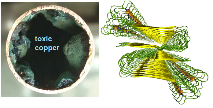
    Copper deficiency and copper toxicity, a dietary conundrum

    A reduced copper diet has been recommended for those at risk for developing Alzheimer’s Disease (Squitti 2014). Others have suggested that the Western diet may not be adequate in copper (Klevay 2011). George Brewer, a colleague of Mitosynergy, has published many papers on Cu2+ in supplements in drinking water, and in residues on produce and their link to Alzheimer’s Disease (Brewer 2015). Dr. Brewer has endorsed Mitosynergy Cu+ supplement.

    A closer look at the gastric bypass surgery

    It is not immediately evident how gastric bypass surgery predisposes a patient for copper deficiency. Some possibilities include

    • improper digestion of copper containing proteins.
    • Copper conjugated peptides are not small enough to be transported by peptide transporters
    • no absorption by Ctr1 and the divalent metal ion transporters in portions of stomach and duodenum that were removed.
    • decreased intake of copper in diet as a result of eating less.
    CuEyeHealth_astricbypass
    The anatomy of a gastric bypass. Ctr1 transporter protein and gene expression from Protein Atlas are included.

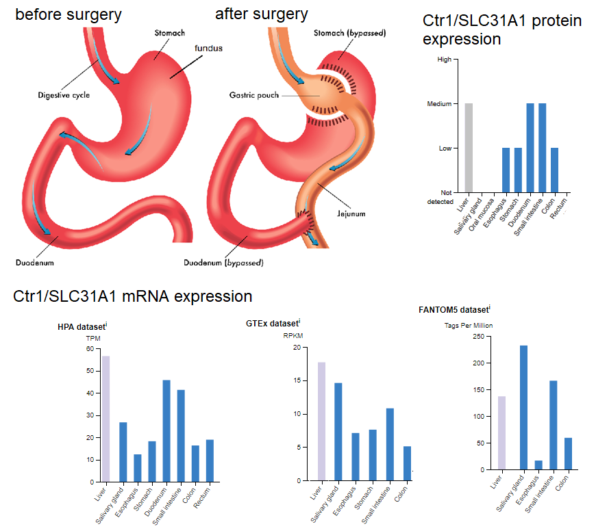
    These possibilities stated, a 1984 study by Urban and coworkers addressed the adaptive responses of the rat small intestine when major portions were removed. Rats were subjected to 50% proximal or 50% distal small bowel resection or sham operation. Four weeks later intestinal absorption of copper was measured in vivo using a recirculation technique. These authors found increases in mucosal mass of non-resected regions as well as increased copper absorption of copper.
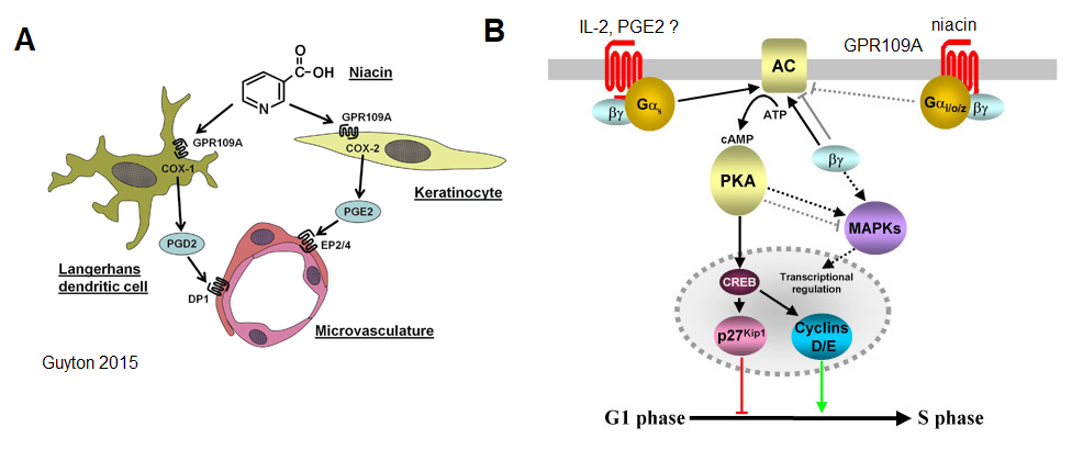
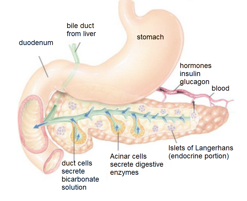
    A preventative therapy for gastric bypass patients is where things get really interesting for Cu(I)NA2. Nicolin and coworkers (2005) demonstrated the presence of the niacin transporting bilitranslocase in the acid secreting parietal cells of the stomach. These cells reside in the fundus, a region that is spared somewhat in the bypass surgery. These authors found the bilitranslocase in the mucous secreting cells of the stomach. This same group found the bilitranslocase in vascular endothelial cells (Maestro 2010 )

    A list of copper containing proteins.
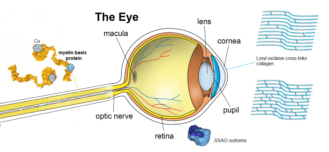
    Myelin

    Myelin is a fatty white substance that surrounds the axon of some nerve cells, forming an electrically insulating layer. This “fatty white substance” also contains a structural protein called myelin basic protein (MBP). In a study of copper deficient rats (Dake 1991), myelin formation was extremely delayed and damaged in the post laminar portion of the optic nerve (Dake 1991). Copper deficiency appeared to cause demyelination or dysmyelination in the optic nerves of rats. MBP is a Cu2+ and 0 Zn2+ binding protein. The of MBP have been studied with electron paramagnetic resonance spectroscopy revealing role of copper in assembly of this important myelin protein (Bund 2010). These same authors noted that the copper chelator cuprizone is used to induce an experimental model of the demyelinating disease multiple sclerosis. As an added note, Dodani and coworkers (2014) performed Cu+ imaging and X-ray fluorescence imaging in the rat developing retina and cultured hippocampus neurons. Disruption by copper chelation or genetic knock down of the CRT1 copper channel altered the spatio temporal properties of spontaneous activity. What role doesCu2+ binding MBP play in buffering this Cu+ activity?

    Monoamine oxidases and SSAO

    The activities of MAO-A, MAO-B and the semicarbazide-sensitive amine oxidase (SSAO) were determined in bovine lens, retina, optic nerve, iris and epithelium. No activities were detected in lens but the SSAO activity was found to be rather evenly distributed in the other tissues (Fernandez de Arriba 1990). SSAO activity towards dopamine was present in the choroid and the retina, but not in the iris or the optic nerve. (Fernández de Arriba 1991). In a later study SSAO, but not its activity, was localized by immunohistochemistry in the human eye (Almulki 2010). SSAO (also known more recently as vascular adhesion protein 1) staining was confined to the vasculature. SSAO labeling showed the highest intensity in both arteries and veins of neuronal tissues: retina and optic nerve, and the lowest intensity in the iris vasculature (p < 0.05). Scleral and choroidal vessels showed moderate staining for SSAO. SSAO intensity was significantly higher in the arteries compared to veins (p < 0.05). More research on the part of Mitosynergy is needed in this area.

    Kaitaniemi (2209) found that the eye version, coded by the AOC2 gene has different substrate specificity than the  general smooth muscle version coded by the AOC3 gene. Protein Atlas data has the expression of AOC2 confined to the retina, though the AOC3 version may be part of the eye and optic verve blood supply.
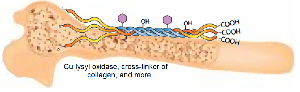
    Lysyl oxidase

    Dudakova and coauthors (2015) published a medical hypothesis that keratoconus, cornea thinning, may be linked to copper deficiency.  Many copper cofactor enzymes have been shown to be altered in keratoconic cornea

    •   superoxide dismutases
    • cytochrome c oxidase
    • lysyl oxidase

    These authors also report a decrease in copper in diseased cornea.

    Some additional copper containing enzymes

    These copper containing enzymes may or may not be important in eye health.

    • L-ascorbate oxidase
    • Superoxide dismutase, Cu/Zn, SOD1 and SOD3.
    • Dopamine beta-monooxygenase
    • Nitrite reductase (NO-forming)
    • Ceruloplasmin (ferroxidase I)
    • Cytochrome c oxidase
    • Dopamine β-hydroxylase
    • Peptidyl glycine alpha-amidating mono-oxygenase (PAM)

    Conclusions

    Gastric bypass patients are sadly at risk of becoming copper deficient and prone to eye problems  We don’t have exact mechanism.  A few possibilities include (1) defects in myelin basic protein, (2) decreased activity of the SSAO monoamine oxidase coded by the AOC2 gene, (3) decreased collagen cross linking performed by lysyl oxidase.  We’ll post more as we learn more.  You may also want to learn more about Copper One Niacin in particular.

    References

    Almulki L, Noda K, Nakao S, Hisatomi T, Thomas KL, Hafezi-Moghadam A.(2010) Localization of vascular adhesion protein-1 (VAP-1) in the human eye. Exp Eye Res.90(1):26-32.
    Bharathselvi M, Biswas S, Raman R, Selvi R, Coral K, Narayanansamy A (2016) Homocysteine & its metabolite homocysteine-thiolactone & deficiency of copper in patients with age related macular degeneration – A pilot study. Indian J Med Res. 2016;143:756–62.

    Brewer GJ (2015) Copper-2 Ingestion, Plus Increased Meat Eating Leading to Increased Copper Absorption, Are Major Factors Behind the Current Epidemic of Alzheimer’s Disease.Nutrients. 7(12):10053-64. Review.

    Bund T, Boggs JM, Harauz G, Hellmann N, Hinderberger D.(2010)Copper uptake induces self-assembly of 18.5 kDa myelin basic protein (MBP). Biophys J. 99(9):3020-8
    Dake Y, Amemiya T.(1991) Electron microscopic study of the optic nerve in copper deficient rats. Exp Eye Res. 52(3):277-81.
    Dodani SC, Firl A, Chan J, Nam CI, Aron AT, Onak CS, Ramos-Torres KM, Paek J, Webster CM, Feller MB, Chang CJ.(2014) Copper is an endogenous modulator of neural circuit spontaneous activity.Proc Natl Acad Sci U S A. 111(46):16280-5.

    Dudakova L, Liskova P, Jirsova K. 2015) Is copper imbalance an environmental factor influencing keratoconus development?. Med Hypotheses. 84(5):518‐524.
    Fernandez de Arriba A, Balsa D, Tipton KF, Unzeta M. (1990)Monoamine oxidase and semicarbazide-sensitive amine oxidase activities in bovine eye.J Neural Transm Suppl.32:327-30.
    Fernández de Arriba A, Lizcano JM, Balsa D, Unzeta M.(1991) Contribution of different amine oxidases to the metabolism of dopamine in bovine retina.Biochem Pharmacol.42(12):2355-61.
    Hirayama Y, Dake Y, Amemiya T.(1992) Cytochrome oxidase in rat ocular tissues with special reference to copper.Acta Histochem.93(1):307-12.
    Kimura E, Hirano T, Yamashita S, Hirai T, Uchida Y, Maeda Y, Uchino M. (2011) Cervical MRI of subacute myelo-optico-neuropathy. Spinal Cord. 49(2):182-5.
    Klevay LM.(2011) Is the Western diet adequate in copper? J Trace Elem Med Biol. 2011 Dec;25(4):204-12. Review.

    Kaitaniemi S., Elovaara H., Groen K., Kidron H., Liukkonen J., Salminen T., Salmi M., Jalkanen S., Elima K. (2009)The unique substrate specificity of human AOC2, a semicarbazide-sensitive amine oxidase. Cell. Mol. Life Sci. 66:2743-2757
    Kumar N, Ahlskog JE, Gross JB Jr. (2004) Acquired hypocupremia after gastric surgery. Clin Gastroenterol Hepatol. 2(12):1074-9.
    Maestro A, Terdoslavich M, Vanzo A, Kuku A, Tramer F, Nicolin V, Micali F, Decorti G, Passamonti S.(2010)Expression of bilitranslocase in the vascular endothelium and its function as a flavonoid transporter.Cardiovasc Res.85(1):175-83

    Moss HE. (2016) Bariatric Surgery and the Neuro-Ophthalmologist. J Neuroophthalmol. 36(1):78-84.
    Naismith RT, Shepherd JB, Weihl CC, Tutlam NT, Cross AH.(2009) Acute and bilateral blindness due to optic neuropathy associated with copper deficiency. Arch Neurol. 66(8):1025-7.
    Nicolin V, Grill V, Micali F, Narducci P, Passamonti S.(2005)Immunolocalisation of bilitranslocase in mucosecretory and parietal cells of the rat gastric mucosa. J Mol Histol. 36(1-2):45-50.
    Pineles SL, Wilson CA, Balcer LJ, Slater R, Galetta SL. (2010) Combined optic neuropathy and myelopathy secondary to copper deficiency. Surv Ophthalmol. 55(4):386-92.
    Pushie MJ, Nienaber KH, Summers KL, Cotelesage JJ, Ponomarenko O, Nichol HK, Pickering IJ, George GN. (2014)The solution structure of the copper clioquinol complex.J Inorg Biochem. 133:50-6.

    Rapoport Y, Lavin PJ. (2016) Nutritional Optic Neuropathy Caused by Copper Deficiency After Bariatric Surgery. J Neuroophthalmol. 36(2):178-81.
    Shah AR, Tamhankar MA.(2014) Optic neuropathy associated with copper deficiency after gastric bypass surgery. Retin Cases Brief Rep.8(1):73-6.
    Urban E, Campbell ME.(1884) Copper absorption by remnant small bowel after extensive intestinal resection in the rat. Am J Clin Nutr. 1984 Sep;40(3):528-35.
    Yarandi SS, Griffith DP, Sharma R, Mohan A, Zhao VM, Ziegler TR.(2014) Optic neuropathy, myelopathy, anemia, and neutropenia caused by acquired copper deficiency after gastric bypass surgery. J Clin Gastroenterol. 48(10):862-5

  • Fatty Liver Disease

    Fatty Liver Disease

    Please note in readying this report on Cu(I)NA2  and fatty liver disease, the authors did not compare Cu(I)NA2  with another dietary copper supplement such as cupric (+2) citrate.  Therefore the conclusions are not unique to Cu(I)NA2. Do not take anything in this post as medical advice. Feel free to discuss this post with your physician.

    This particular study came out of the Department of Physiology of Aswan University, in Aswan, Egypt [1].  The Liver Foundation estimates that approximately 30% of the U.S. population suffers from non alcoholic fatty liver disease and 5% are afflicted by  its subtype nonalcoholic steatohepatitis.  These numbers translate to about 100 million individuals in the United States living with  nonalcoholic fatty liver disease.  The global prevalence of fatty liver disease is  about as high.  Hegazy and coworkers were not interested in the mechanisms of moving fats out of the liver and into the blood stream.  They simply wanted to know if Cu(I)NA2 might relieve the inflammation associated with fatty liver disease.  Copper might have a role in both.

    Fatty liver disease

    Fatty liver disease is, very simply, the abnormal accumulation of fats in the liver.  Alcoholism is a main cause of fatty liver disease.  Non alcoholic fatty liver disease my be caused dietary deficiencies, metabolic abnormalities, drugs and toxins, and immune responses. These  authors chose to induce FLD in  rats with a methionine- and  choline deficient diet (MCDD).

    What are methionine and choline?

    Methionine is an essential amino acid that participates in many enzymatic reactions as a methyl donor.  DNA methylation is one of many of these reactions.  Note the methyl group in Figure 1c.  Choline (Fig 1c) is the basic constitute of lecithin (phosphatidyl choline), a phospholipid found in plant and animal cells.  Choline may also serve as a methyl donor.  Hydrogens in the structures in Figure 1c are “understood.”  The end of the sticks are understood to be methyl groups (-CH3).  Dietary deficiency choline may result in accumulation of fat in the liver due to lack of very low density lipoprotein (VLDL) needed to transport fats out of the liver.  Methionine deficiency may cause general liver damage that may be clinically measured by the release of the liver enzyme alanine amino transferase (ALT) into the blood.    PubChem tells us that choline may be used to synthesize betaine.

    Choline deficiency in dairy cattle transitioning from being pregnant to being milk producers is an industry concern [2].  One of the industry challenges is protecting the dietary choline from the contents of the rumen.

    FLD_1
    Figure 1 The intersection of a methionine [1] and b choline pathways.  c a structure of methionine and chloine. d In addition to B12, methyl synthase has Mg2+ cofactors.

    Why Copper?

    A 1999 study examined the influence of copper deficiency in rats on folate and homocysteine synthesis.  Hepatic folate, and plasma vitamin B-12 concentrations were similar in both groups [3].  Homocysteine in the blood plasma increased, most likely the result of a decrease in hepatic methionine synthase (MS) activity [3].  The authors speculated that MS might be a cuproenzyme in addition to requiring B12 cofactor.  Twenty years later, we still have no clue.  Rat methionine synthase has three amino acids that interact with  Mg2+ and two that interact with K+ these were found by following the UniProt line to  an X-ray crystal structure of methionine synthase. [4]

    FLD_2
    Figure 2 Interaction of amino acids in methionine synthase with ATP and free methionine , adapted from [4]

    One would think that if Cu2+  can substitute for Mg2+  in methionine synthase, we’d not know it by now.  Mg has only one oxidation state.   Cu has two.  The implications on the catalytic process would be interesting, if such a substitution were the case. The answer may lie in the affect of Cu on gene expression.

    Cu(I)NA2 protects the fatty liver from further damage

    FLD_3
    Figure 3 from [1]  Effect of CNC, Cu(I)NA2, on liver enzyme activity in the serum of rats with fatty liver (mean±standard deviation, n=10).Means with different superscripts in the same row are significantly different at p<0.05. CNC=Copper-nicotinate complex, MCDD=Methionine- and choline-deficient diet, ALT=Alanine aminotransferase, AST=Aspartate aminotransferase, GGT=Gamma glutamyl transferase

    Note that CNC,  Cu(I)NA2, brings the activities of liver in the serum down to control levels even in the presence of methionine and choline deficiency.

    FLD_4
    Figure 4 from [1]  Effect of CNC on oxidative/anti-oxidative markers in liver homogenate of rats with fatty liver (mean±standard deviation, n=10).  Means with different superscripts in the same row are significantly different at p<0.05. CNC=Copper-nicotinate complex, MCDD=Methionine- and choline-deficient diet

    Note that Cu(I)NA2, brings reduced glutathione to control levels in the fatty liver model.    Malondialdehyde, a marker of reactive oxygen species degradation of polyunsaturated fatty acids,   is  decreased to control levels. Cu(I)NA2 almost doubles superoxide dismutase activity [1].  The authors did not distinguish between superoxide mimetic activity of  Cu(I)NA2  alone or in intracellular Cu/Zn SOD1 or extracellular Cu/Zn SOD3 [1].   These authors also looked at expression at various cytokines associated with inflammation.

    Copper regulates gene transcription

    Liver gene expression was examined in a “tx-j” mouse model of Wilson’s Disease caused by a mutation in ATP7B.  ATP7B secretes excess copper into the bile.  Non functional in WD patients have a toxic overload of copper in their livers.  Shibata and  coauthors [5] looked at gene expression (Y-axis, Fig 5A) for many stages of development.  They were particularly interested in genes that regulate DNA methylation.  We will stick to our story line and single out two genes that might impact fatty liver disease.

    FLD_6
    Figure 5 How copper might regulate fatty liver disease A. Relative expression between two select genes in control mice and a mouse model of Wilson’s disease B. Re-visitation of Figure 1 with gene expression data

    We have no way of knowing if dietary copper of any sort mimics the effect of ATP7B defect (tx-j) copper overload.  In such a hypothetical methioinine synthase compensates for reduced dietary methione and possibly even choline.  By most accounts, S-adenosyl homocyetinase merely speeds up the equilibrium between homocysteine and S-adenosyl homocysteine.  The expression of this gene is decreased by copper overload.

    Could Cu(I)NA2  regulate gene expression in the fatty liver in a manner that facilitates fat export?  We do not know! A certain amount of caution needs to be used that genes are over-expressed when there is too much copper are not expressed enough when there is copper deficiency.

    Concluding remarks

    •   This featured Cu(I)NA2 study [1] was not concerned with enzymes involved with fatty liver disease.  The lessened liver damage and positive oxidative status results are the encouraging focus of this report.
    • Dietary deficiency in choline/methionine can impact can lead to fatty liver disease in dairy cattle [2].
    • Earlier rodent studies suggest a link between copper and the methionine cycle [3] .  Twenty years later there is no evidence that copper is a cofactor in methionine synthase.  Considering how magnesium does fit into the structure [4], it would be interesting if it did.
    • The most likely explanation for Reference [3] data is copper regulation of hepatic gene transcription, in particular methione synthase [5].
    • We want to emphasize that we are not making medical claims regarding Cu(I)NA2 in this post.  
    • We do see enough data to support investigative studies.

    References

    1. Hegazy AM, Farid AS, Hafez AS, Eid RM, Nasr SM. (2019) Hepatoprotective and immunomodulatory effects of copper-nicotinate complex against fatty liver in rat model. Vet World. 12(12):1903-1910. [PMC free article]
    2. Abbasi, I.H.R., Abbasi, F., Soomro, R.N. et al. Considering choline as methionine precursor, lipoproteins transporter, hepatic promoter and antioxidant agent in dairy cows.(2017) AMB Expr 7, 214 (2017). [Cross Ref]
    3. Tamura T, Hong KH, Mizuno Y, Johnston KE, Keen CL. (1999) Folate and homocysteine metabolism in copper-deficient rats. Biochim Biophys Acta. 1427(3):351-6.
    4. González B, Pajares MA, Hermoso JA, Guillerm D, Guillerm G, Sanz-Aparicio J (2003) Crystal structures of methionine adenosyltransferase complexed with substrates and products reveal the methionine-ATP recognition and give insights into the catalytic mechanism. J. Mol. Biol. 331 407-16
    5. Le A, Shibata NM, French SW, Kim K, Kharbanda KK, Islam MS, LaSalle JM, Halsted CH, Keen CL, (2014)Characterization of timed changes in hepatic copper concentrations, methionine metabolism, gene expression, and global DNA methylation in the Jackson toxic milk mouse model of Wilson disease. Medici V. Int J Mol Sci. 2014 May 7;15(5):8004-23. [Cross Ref]
  • minerals and bone health

    minerals and bone health

    The osteon is the basic unit of compact bone.

    BoneHealth_osteon
    Figure 1, an osteon, adapted from lecturio.com


    Mineral wheels, aka Mulder charts

    A Mulder’s chart, also known as a “mineral wheel” are often used in agriculture circles to describe mineral interactions.  Variations of this wheel are all over the Internet with no one (including us) citing the original origin.   Because these representations are “all over the place” they make good places to start a discussion.


    BoneHealthMineralInteraction
    Figure 1 A Mulder chart/mineral wheel showing mineral interactions. Strontium and silicon, while important for bone health are not in most representations on the Internet.

    Calcium and phosphate are both important in bone health.  Cola, but not other carbonated beverage consumption, was found to be associated with lower bone mineral density in women, but not men (Tucker 2006).   While this site is mostly about copper, other minerals may deserve mention.

    Bone is hydroxylapatite

    a fancy name for Ca10(PO4)6(OH)2

    10Ca(OH)2 + 6H2PO4 +2H2O → Ca10(PO4)6(OH)2 + 14H+

    In examining this equation it becomes very clear that making bone, hydroxylapatite, from calcium and phosphate H2PO4 ) can become a complicated matter of where to put the 14H+. This is an acid balance issue.

    Calcium, signaling and precipitation with phosphate

    • The cells of bone tissue control over 99% of the human body’s calcium content, though not calcium sensors themselves.
    • The principal calcium sensors that regulate bone calcium flux are located in the parathyroid glands.
    • Bone function is also modified by vitamin D and by calcium transport in the kidney and intestine.
    • This massive amount of transport is carried out by specialized transporters, not only for calcium, but also for other minerals needed to maintain charge balance. (Blair 2007)


    BoneHealthOsteoblast
    Figure 2, adapted from Blair (2007) figure 1. Osteoblasts, on the surface of the bone,  are connected into sheets of cells  by gap junctions containing connexin-43.  .

    Pyrophosphate is derived from ATP. Everything is connected by structures called gap junctions. The osteoblasts secrete an organic matrix comprised mainly of type I collagen and other proteins (Blair 2007).

    • type I collagen,
    • small calcium binding protein osteocalcin, which facilitate mineral deposition
    • alkaline phosphatase activity which degrades pyrophosphate
    • pyrophosphate membrane transporters such as  ANKH,


    Osteoclast degrade bone

    BoneHealthOsteoclast
    Figure 3, adapted from Blair (2007) and other images on the Internet. Cathepsin K and matrix metalloproteas (mmp) are proteases that degrade the collagen component of bone.


    Copper!

    We’ve covered that we have collagen in our bones.  Lysyl oxidase are a family of copper cofactor enzymes that cross-link collagen.  Copper deficiency is thought to impact bone health via lsyl oxidase (Medeiros 2016).  In a rodent model mechanical unloading was found to increase reactive oxygen species as well as increase the expression of Cu/Zn SOD1.  SOD1 deficient mice experienced greater bone loss.  Did you know that collagen in our corneas and that copper is necessary for eye health? Some think that cuprous forms of copper may be the best way of getting that copper. .

    Manganese

    • Many websites give the content of manganese as 10-20mg.  Wikipedia gives a number of 12 mg.  for a normal adult human with most being in the bone followed by the liver, and the kidney. It was not clear if bone is just a storage organ with the liver and kidney being sites of accelerated metabolism.
    • Manganese induced ricketts is associated with phosphate depletion.  (Svensson 1987)
    • Calcium and parathyroid hormone increased in the sera o f manganese deficient chicks.  (Zhaojun 2013)  PTH stimulates bone resorption.

    Landete-Castillejos (2012) formed a hypothesis on the role of manganese in bone from field observations.

    • Antler bone, one of the toughest of all bones was chosen as a model
    • A cold winter in Spain led to decreased Mn content of plants and ultimately a decreased Mn concentration in Antlers.
    • These antlers had decreased mechanical strength.
    • This observation led to the hypothesis called “essential mineral for calcium fixation” (EMCaF)
    • A diet depleted from Mn but not Ca produced femurs with levels lower in Mn and Ca in rats that were growing at a normal rate with no other effects of the diet. (Landete-Castillejos 2012)
    • An image search on “manganese and phosphate” was used to find a compound called purpurite.
    • On the other hand, the UniProt database contains numerous human Mn Proteins.  of which the manganese superoxide dismutase 2 (SOD2) is one.
    • UnitProt also has even more plant Mn Proteins.  The sweet potato purple acid phosphatase probably has the most interesting sounding name.  This protein uses a aspartic acidic, an asparagine, and two histidines to coordinate manganese.

    BoneHealth_manganese
    Figure 4.  Manganese complexes A. with phosphate, aka purpurite B. with SOD2, UnitProt.org  C. the sequence of SOD2 with the Mn binding sites noted with orange boxes. H, histdine, D, aspartic acid.

    What does manganese do in bone?  It could form complexes with the phosphate or it could form complexes histidines and other amino acids in collagen.  CuCl2  is plant copper.  MnPO4  is plant manganese.  In the opinion of Mitosynergy, minerals for humans should come as organic complexes,.

    Magnesium, technology imitating nature

    Magnesium and magnesium oxide have become favorite doping agents for synthetic hydroxyl apatite used for bone rebuilding and other tissue engineer applications. Magnesium is important for cell-extracellular matrix interactions and bone apatite structure and density (Hickey 2015).

    Strontium

    These are some tidbits when we first started looking at bone minerals a few years ago.

    • Strontium ranelate increases the proliferation of pre-oseoblasts (make hydroxyapetite) as well as the production of collagen. (Fonseca 2008)
    • In addition to the osteoporosis approved drug strontium ranelate, Sr fructose, 1,6-diphosphate, and strontium citrate have also proven effective in treating osteoporosis. (Tan 2014)

    A Marx (2020) review seeks to add clarity to the confusion of what this element in the same periodic group as calcium is doing in bone.

    • The Ca-sensing receptor (CaSR)  that regulates parathroid hormone senses strontium as well.
    • CaSR is in osteoblasts
    • CaSr is in osteoclasts
    • In mineralization, Sr substitutes for Ca in hydroxy apatite.
    • Sr interacts with collagen
    • Sr can coordinate with 8 oxygens

    We are showing the sequence of one of the repeats of collagen 1.

    Note that collagen may also be glycosylated with polysaccharides, hexagons in figure 5B.

    BoneHealth_strontium
    Figure 5 Strontium in bone health  A Marx proposed oxides of strontium bind to collagen fibres.  B.  Collagen fibers exist as triple helices with repeating sequences.  One such repeat is shown.  Glutamic acids are noted with orange boxes  C.  the SER sequence shown in panel B might bind phosphate and calcium or strontium.

    Marx (2020) claim that hydroxyapatite precipitates on collagen fibers (Fig 5A).  How?  Figure 5B shows one repeat of one of three giant monomers in a collagen fiber.  Note the glutamic acids (E).  We also need to remember that collagen fibers are glycosylated.  Figure 5C shows the structure of a Serine-glutamic acid-arginine peptide.  Yeah, it kind of makes sense that hydroxy apatite might precipitate onto collagen.  Strontium is in the same periodic group as calcium.  How its larger atomic radius makes a difference in bone health is beyond the scope of this post.

    Silicon

    The  Jugdaohsingh (2007) review was interesting but hard to visualize.  Here are dome key points:

    1. Si(OH)4 interacts readily with alkyl diols of sugars to form five and six-coordinate Si complexes suggesting that interactions with bio-molecules is possible. Are these similar to borate interactions with vicinal cis-diols?
    2. Plants produce biogenic (phytolithic) silica which is often associated with the polysaccharide/carbohydrate components of the cell wall
    3. Monocot grains generally accumulate 10-20x as much silica as dicot legumes.
    1. Orthosilicic acid is thought to be the breakdown of phytosillicates that actually gets absorbed.
    2. In the rat highest levels or orthosilicates found in bone and other connective tissues such as, skin, nail, hair, trachea, tendons and aorta and very much less (10-20 fold less) in soft tissues
    3. Orthosilicic acid have also reported increases in type I collagen synthesis and cellular differentiation
    4. High levels of Si were found to be strongly bound to connective tissues and its components, namely glycoaminoglycans, polysaccharides and mucopolysaccharides.
    5. Silicon has also been found at the mineralization front of growing bone


    Bhattacharjee H, Mukhopadhyay R, Thiyagarajan S, Rosen BP (2008) Aquaglyceroporins: ancient channels for metalloids J Biol. 2008; 7(9): 33.

    BoneHealth_silica
    Figure 6.  Silicate forms bonds with molecules with hydroxyl groups A. Periodic table of elements. Inset contains image of silicate acid B. Silica complexing with vicinal hydroxyl groups of two mannose molecules.

    Mannose is a six carbon sugar present in polysaccharides of the extracellular martix.   Lysyl oxidase increases bone strength by cross-linking lysine residues in collagen.  Any secreted protein or surface protein that is glycosylated with mannose or other sugars may be cross-linked by silica.

    Borate/Boron

    Nielsen (2016) reviewed some of the physiological functions of borate

    1.  Boron binds nicotinamide adenine dinucleotide (NAD+) with high affinity.  Whether boron acts on intracelular and/or extracellular NAD+was not that clear in this review.
    2. One role of extracellular NAD+ is binding to the plasma membrane receptor CD38, an adenosine diphosphate ribosyl cyclase that converts NAD+ to cyclic ADP ribose.
    3. Cyclic ADP ribose is released intracellularly and binds the ryandodine receptor,which induces the release of calcium ions from the endoplasmic reticulum.
    4. Cell culture studies show that boron binds to and is a reversible inhibitor of cyclic ADP ribose
    5. Boron in concentrations that are found in blood was found to decrease Ca2+ release from ryandodine receptor-sensitive stores.
    6. It has been hypothesized that boron is bioactive through binding NAD+ and/or cyclic ADP ribose and inhibiting the release of Ca2+, which is a signal ion for many processes affected by boron, including insulin release, bone formation, immune response, and brain function.

    Forrest Nielsen has done a lot of work in bone and tooth strength in rabbits with boron supplementation. There’s also the component of high energy diet. To be honest, borate binds to so many compounds with cis-vicinal hydroxyl groups, it’s kind of hard to tell which of many possible interactions are the important ones.  Another review, Park (2006) covered boron in plants and animals.  In reviewing both of these reports it is important to remember that borate binds carbohydrates

    BoneHealth_boron
    Figure 7 Boron in bone health A. Structure of NADH and NAD+ . A red box is drawn around one of the ribose sugars. B. Borate binding to ribose C. The borate transporter described by Park (2006)

    • Boron stabilizes NADH oxidase in plants
    • In mammalian tissue too. Highest concentrations are in bone, heart, spleen, and liver.
    • Required for a hydroxylation step in the synthesis of testosterone, and 17-β-estradiol.
    • Bor1 and NaBC1 are plant and mammalian homologs of the same transporter.
    • In a study of cultured cells, 0.1 to 1mM borate promoted proliferation. 10mM borate was cytotoxic.

    Conclusion

    Note that all of the minerals covered in this bone health post bind to organics.  Bone is very much an organic material in addition to the hydroxy apatite most of us learned in school.    Perhaps we need to throw that mineral wheel when it comes to mineral supplements for humans.  As of this posting, Mitosynergy only sells cuprous niacin, not the other minerals.    Thank you for reading!

    References

    Bhattacharjee H, Mukhopadhyay R, Thiyagarajan S, Rosen BP (2008) Aquaglyceroporins: ancient channels for metalloids J Biol. 2008; 7(9): 33.

    Blair HC, Schlesinger PH, Huang CL, Zaidi M.(2007)Calcium signaling and calcium transport in bone disease. Subcell Biochem.45:539-62. Review.

    Fonseca JE (2008) Rebalancing bone turnover in favour of formation with strontium ranelate: implications for bone strength Rheumatology (Oxford) 2008 July; 47(Suppl 4): iv17–iv19.

    Kim MH, Bae YJ, Choi MK, Chung YS.(2009) Silicon supplementation improves the bone mineral density of calcium-deficient ovariectomized rats by reducing bone resorption. Biol Trace Elem Res. 2009 Jun;128(3):239-47.

    Hickey DJ, Ercan B, Sun L, Webster TJ.(2015) Adding MgO nanoparticles to hydroxyapatite-PLLA nanocomposites for improved bone tissue engineering applications. Acta Biomater. 14:175-84.

    Jugdaohsingh R (2007) Silicon and bone health J Nutr Health Aging. 11(2): 99–110.

    Landete-Castillejos T, Molina-Quilez I, Estevez JA, Ceacero F, Garcia AJ, Gallego L. (2012)
    Alternative hypothesis for the origin of osteoporosis: the role of Mn. Front Biosci (Elite Ed). 2012 Jan 1;4:1385-90.

    Marx D, Rahimnejad Yazdi A, Papini M, Towler M. (2020) A review of the latest insights into the mechanism of action of strontium in bone. Bone Rep. 2020 Apr 24;12:100273.

    Medeiros DM. (2016) Copper, iron, and selenium dietary deficiencies negatively impact skeletal integrity: A review. Exp Biol Med (Maywood). 2016 Jun;241(12):1316-22

    Morikawa D, Nojiri H, Saita Y, Kobayashi K, Watanabe K, Ozawa Y, Koike M, Asou Y, Takaku T, Kaneko K, Shimizu T. (2013) Cytoplasmic reactive oxygen species and SOD1 regulate bone mass during mechanical unloading. J Bone Miner Res. 2013 Nov;28(11):2368-80

    Nielsen FH (2014) Update on human health effects of boron. J Trace Elem Med Biol.28(4):383-7..

    Park M, Li Q, Shcheynikov N, Muallem S, Zeng W.(2005) Borate transport and cell growth and proliferation. Not only in plants. Cell Cycle. 4(1):24-6.

    Svensson O, Engfeldt B, Reinholt FP, Hjerpe A.(1987) Manganese rickets. A biochemical and stereologic study with special reference to the effect of phosphate. Clin Orthop Relat Res. 1987 May;(218):302-11.

    Tan S, Zhang B, Zhu X, Ao P, Guo H, Yi W, Zhou GQ.(2014)Deregulation of bone forming cells in bone diseases and anabolic effects of strontium-containing agents and biomaterials.Biomed Res Int. 2014:814057

    Tucker KL, Morita K, Qiao N, Hannan MT, Cupples LA, Kiel DP.(206)Colas, but not other carbonated beverages, are associated with low bone mineral density in older women: The Framingham Osteoporosis Study. Am J Clin Nutr. 2006 Oct;84(4):936-42. doi: 10.1093/ajcn/84.4.936.

    Zhaojun W, Lin W, Zhenyong W, Jian W, Ran L.(2013) Effects of manganese deficiency on serum hormones and biochemical markers of bone metabolism in chicks. J Bone Miner Metab. 31(3):285-92

  • Niacin benefits infections

    Niacin benefits infections

    Our featured image is a reminder that the niacin receptor is a member of the G protein coupled receptor family.  These receptors talk to each other via α subunits.

    Niacin is the other two thirds of Cu(I)NA2., a cuprous niacin supplement available on the market.  Most healthcare providers are probably aware of niacin’s many functions.

    A big unknown for us is if Cu(I)NA2 can even bind to the niacin receptor.  If nothing else, the other two thirds modulate the immune system.

    1. Niacin is a  precursor to NAD/NADH2, an H+ / electron carrier in numerous biochemical reactions.
    2. Niacin in high doses causes  dilation of the skin’s vasculature in a  response called “flush.”
    3. High doses of niacin are available in prescription form as Niaspan® and its generic equivalent “niacin ER” to lower  low density lipoprotein (LDL) cholesterol

    Niacin receptors in neutrophils and more

    Apart from being a precursor of NADH2, niacin has direct physiological actions on its  high and low affinity receptors

    1. NIACR1 is also known as the hydroxy carboxylic acid receptor (HCA2) GPR109A, HM74a, HM74b.NIACR2.
    2. NIACR2 is also known as HCA3 and GPR109B.  It is the low affinity receptor that is activated by amounts of niacin not normally found in the diet.

    Protein Atlas has compiled some nice data on the mRNA expression of GPR109A in blood cells.

    NiacinInfections1
    Top. Consensus mRNA expression of GPR109A, a niacin receptor, in blood cells. Bottom Data for dendritic cells, microglia, and macrophage are harder to obtain when they migrate into the tissue site of an infection. GPR109A expression in macrophage may be induced by lipopolysaccharide (LPS) from Gram negative bacteria.

    Niacin receptors and cAMP

    A 2008 study of Kostylina demonstrated that niacin promotes apoptosis, programmed cell death,  in mature but not immature neutrophils.  This action is mediated by the GPR109A G-protein coupled receptor.  G-protein coupled receptors may stimulate or inhibit the enzyme adenylyl cyclase.  AC converts cellular ATP to the second messenger cyclic AMP (cAMP).  Note the cyan colored  βγ subunit.  These will come into the discussion of potential niacin signalling in macrophage.

    NiacinInfections2
    A Niacin GPR109A pathways to vasodilation seen in “flush” B G-protein coupled receptors responsible for pro-survival pathways in neutrophils couple to Gs that activates adenylyl cyclase. The niacin receptor GPR109A couples with Gi that inhibits adenylyl cyclase (AC).

    G protein coupled receptor cross talk

    The authors did not discuss which G-protein coupled receptor on neutrophils might be simulating adenylyl cyclase by way of a Gs subunit.  The interleukin-2 and PGE2 receptors are G-protein coupled receptors that may stimulate neutrophils.

    Niacin, GPR109A, and macrophages

    Inflammatory mediators LPS ( Gram negative bacteria), zymosan (polysaccharide in  yeast, fungi),  lipoteichoic acid (LTA, Gram positive bacteria), polyinosine-polycytidylic acid (poly I:C, viruses) induce GRP109A in cultured marcrophage.  (Feingold 2014).   LPS was able to increase GPR109A mRNA up to 80x in less than 24 hours.  Niacin binding to GRP109A receptor may be a means of turning off the initial, innate immune response to pathogens.

    Niacin inhibits macrophages by decreasing cAMP

    Zandi-Nejad and coworkers (2013) examined the role of niacin on macrophage function.  Their primary focus was the role of macrophage in atherosclerosis rather than infectious diseases.  The”cytokine storm“, also known as cytokine release syndrome, has gained a lot of attention as a morbidity factor in COVID-19.

    1. Phagocytosis and chemotaxis  of blood wild type HCAR2+/+  and  HCAR2-/-   knock out mouse bone marrow derived marcophage (BBM) was shown to be decreased by nicotinic acid.  The HCAR2 gene codes for the GRP9A protein.
    2. LPS induction of cytokines TNFα, IL-6, IL-12p40, and IL-1β was measured in
    3. HCAR2+/+  and  HCAR2-/-   BBM.  Niacin lowered mRNA transcripts of all of these cytokines after 48 hours of exposure to LPS in the wild type but not in the BBM lacking the GPR109A receptors.

    Zandi-Nejad and coworkers (2013) remarked that their results differed from those of Rossi (1998) in that cAMP increases inhibited macrophage.  Shi (2017) suggested that niacin macrophage inhibition had more to do with the βγ subunit of heterotrimeric G proteins acting on protein kinase C.

    A look back, cAMP and macrophage activity

    Rossi (1998) demonstrated that intracellular cAMP decreased the ability of preipheral blood monocyte derived macrophage to phagocytize apoptotic neutrophils but not opsonized red blood cells.  These  authors also found that PGE2 to be a strong inducer of cAMP.

    A look ahead at recent publications

    Negreiros-Lima and coworkers (2020) have pushed the boundaries of earlier studies.  These authors used a non-hydrolyzable analog of cAMP, db-cAMP to activate protein kinase A pathways.  They addressed ways that cAMP modulates the M1 (pro inflammatory) to M2 (anti-inflammatory) transition.  To make matters more complicated, they introduced the M2 subsets: M2a, M2b and M2c.   We have no idea if the GRP109A receptor is expressed in all of these developmental stages.  An additional new term is  “effectocytosis” the clearance of neutrophils undergoing apoptosis.

    References

    Bühler S, Frahm J, Liermann W, Tienken R, Kersten S, Meyer U, Huber K, Dänicke S.(2018) Effects of energy supply and nicotinic acid supplementation on phagocytosis and ROS production of blood immune cells of periparturient primi- and pluriparous dairy cows. Res Vet Sci.116:62-71

    Guyton J., Campbell K., Lakey W. (2015) Niacin: Risk Benefits and Role in Treating Dyslipidemias. In: Garg A. (eds) Dyslipidemias. Contemporary Endocrinology. Humana Press, Totowa, NJ

    Feingold KR, Moser A, Shigenaga JK, Grunfeld C.(2014) Inflammation stimulates niacin receptor (GPR109A/HCA2) expression in adipose tissue and macrophages. J Lipid Res. 2014 Dec;55(12):2501-8. Link

    Kostylina G, Simon D, Fey MF, Yousefi S, Simon HU.(2008) Neutrophil apoptosis mediated by nicotinic acid receptors (GPR109A). Cell Death Differ.15(1):134-42  Link

    Negreiros-Lima GL, Lima KM, Moreira IZ, Jardim BLO, Vago JP, Galvão I, Teixeira LCR, Pinho V, Teixeira MM, Sugimoto MA, Sousa LP. (2020) Cyclic AMP Regulates Key Features of Macrophages via PKA: Recruitment, Reprogramming and Efferocytosis. Cells. 9(1). pii: E128. Link

    Rossi AG, McCutcheon JC, Roy N, Chilvers ER, Haslett C, Dransfield I. (1998) Regulation of macrophage phagocytosis of apoptotic cells by cAMP. J Immunol. 160(7):3562-8.Link

    Shi Y, Lai X, Ye L, Chen K, Cao Z, Gong W, Jin L, Wang C, Liu M, Liao Y, Wang JM, Zhou N. (2017) Activated niacin receptor HCA2 inhibits chemoattractant-mediated macrophage migration via Gβγ/PKC/ERK1/2 pathway and heterologous receptor desensitization. Sci Rep. 2017 Feb 10;7:42279 Link

    Zandi-Nejad K, Takakura A, Jurewicz M, Chandraker AK, Offermanns S, Mount D, Abdi R (2013) The role of HCA2 (GPR109A) in regulating macrophage function. FASEB J. 27(11):4366-74. Link

  • safe copper for seniors

    safe copper for seniors

    Proper Cu+ and Zn2+ absorption

    2021 update: This post has been password protected since it was created May 19th 2020.  You may not determine whether or not copper is safe for you or a senior you love by reading this post.  You are cordially invited to take this information to your physician and have a discussion with them as to whether Cu+ is safe based on the peer reviewed science presented in this post. 

    Some health care providers have expressed concern that copper in the diet may interfere with zinc absorption.  Based on a recent Maares and Haase (2020) review, it is hard to conceive how Cu+ could interfere with Zn2+  absorption by  Zip4 in the apical membrane and excretion from the enterocyte into the blood by  ZnTn-1.   Perhaps what Mitosynergy calls toxic copper, Cu2+ , could conceivably interfere with Zn2+  absorption.  Doguer (2018) and Maares and Haase (2020) mention the possibility of Zn2+ and Cu2+  entering the enterocyte by  way of a divalent metal ion transporter.  Doguer (2018)  has labeled the reductase in proximity to Ctr1 as a brush-border membrane (BBM) ferric iron reductase duodenal cytochrome B (DCYTB) that can reduce +3  iron and  +2  copper. These include the major iron importer divalent metal-ion transporter 1 (DMT1).  

    CopperSeniors2
    Copper in the +1 oxidation state is absorbed by distinct pathways from that for zinc.

    Clinicians might also enjoy a Nishitio and Kambe (2018) review on copper, zinc, and iron.absorption from the gut.  Whether DCYTB is a  Cu2+  reductase for Ctr1 seems to remain a question.    One could argue that the Mitosynergy’s  Cu(I)NA2  is a specially formulated copper supplement that does not require this reductase.

    Copper and Immune Function

    • Copper and the immune system started out with an examination of the role of the neutrophil to lymphocyte ratio that was reported to be out of whack in severe COVID-19 infections. Don’t we need neutrophils as part of our innate immune system? Peer reviewed literature was reviewed on the impact of copper deficiency on the innate and antibody producing adaptive immune system.
    • Fish and Human Studies is a presentation of data on Cu(I)NA2 on neutrophil and lymphocyte counts in fish and humans.    improves antibody production against a fish pathogen in the fish studies.  Some thoughts on the role of Cu(I)NA2 in beefing up the mitochondria in antibody producing plasma cells are presented.
    • Copper and the renin agiotensin system  Hypertension is a risk factor for severe COVID-19 infections.  This virus binds to the angiotensin converting enzyme 2 (ACE2) cell surface enzyme.  ACE2 is part of the renin-angiotensin-system (RAS) that our body uses to maintain blood pressure.  It is activated by circulatory collapse that may occur in bacterial and fungal infections.  Activation of the RAS is often associated with super oxide, a reactive oxygen species, production.  I looked at literature showing that angiotensin II may up regulate the expression of Cu/Zn superoxided dismuatase 3.  

    Proper Copper and the Copper-2 Hypothesis

    Dr George Brewer is a professor of Human Genetics at the University of Michigan.  Dr Brewer became interested in the link between Alzheimer’s Disease and copper in the +2 oxidation state from water pipes in his studies of Wilson’s Disease.  Wilson’s Disease patients have a defect in the copper export protein ATP7B that prevents them from eliminating excess copper in the bile, and ultimately the feces.  Dr. was investigating tap water from sites across the United States.  He found that tap water from many locals had Cu2+ exceeding concentrations found to exacerbate progression of Alzheimer’s Disease in animal models.  Dr Brewer is hypothesizing that Cu2+ does not cause Alzheimer’s Disease but rather enables the epidemic (Brewer 2019).  Dr Brewer also suggested that Cu2+ found in most dietary supplements could hasten the progression of Alzheimer’s Disease.  The historical increase in meat consumption was down played as source of increased dietary copper.  Dr Brewer hinted at the importance of two pools of blood borne copper:  (1) bound to ceruloplasmin and (2) “freely” bound to blood proteins like albumin.

    Dr Marc Solioz of Marc Solioz Department Clinical Research, University of Bern, dismisses the Copper-2 hypothesis.  The United State and Switzerland have about the same incidence of Alzheimer’s Disease even though the U.S population consumes more red meat, takes more supplements, and is more likely to have copper pipes.  Switzerland is unique in that copper water pipes were never used.

    Ceruloplasmin bound copper and free copper

    Techio and others (2016) of the BioMarkers Laboratory of Brescia, Italy found that non-ceruloplasmin bound plasma copper correlated with Alzheimer’s Disease brain abnormalities compared with ceruloplasmin bound copper. Squitti and coworkres (2014) followed patients with mild cognitive impairment (MCI) to test the hypothesis that non-ceruloplasmin bound copper is a risk factor for progression to ALzheimer’s Disease.  They concluded that their results might warrant dietary intervention to lower the risks.  Park and coworkers (2013) found elevated copper and ceruloplasmin in Korean Alzheimer’s Disease patients.  An inverse relationship was found between cognitive performance and serum copper.  These authors also speculated that Alzheimer’s Disease patients may have defective (apo) ceruloplasmin that lacks the copper cofactor.  Wang and Wang (2019) in their review argue that properly functioning ceruloplasmin is neuroprotective.

    A note of skepticism

      George Brewer and others make a very compelling argument for Cu(II) being toxic copper.  According to conventional wisdom reviewed by Doguer (2018) Cu+ is secreted directly into the portal circulation where it is picked up by the liver for packaging into ceruloplasmin.  Surely Cu+ would not remain in the +1 oxidation state in this unchaperoned mode. Most models have the Menkes ATPase (ATP7A) directly secreting Cu+ into the blood.  

    • Are the oxygen tensions in the portal vein low enough to preserve the +1 oxidation state long enough to get to the liver?
    • ATP7A loads SOD3 in the Golgi.  Is there any indication of regulated release of  Cu+ to apo copper binding proteins in the serum?   An abstract from a Russian language publication (Tsymbalenko 2000) synthesized peptides corresponding to two of the four extracellular loops of ATP7A.  One of these peptides was able to bind to a domain of the blood copper transport protein ceruloplamin.

    The following was obtained from the UniProt.org database.

    CopperSeniors3
    The first extracellular loop of ATP7A has histidines and other amino acids that could aid in the coordinated transfer of Cu(I) to target blood proteins.

    Concluding Remarks

    The manner in which the COVID-19 has hit nursing homes and the elderly hard has lead Mitosynergy to reexamine old data on Cu(I)NA2  and immune function.  We also have to reexamine the “copper-2 hypothesis” regarding dietary copper and general fear that copper in any oxidation state potentiates Alzheimer’s Disease.  The general consensus is that ceruloplasmin bound copper is okay and it is the “free” copper bound non specifically to serum proteins that is an Alzheimer’s Disease risk factor.    We can propose a mechanism by which  dietary Cu+  is more likely to end up ceruloplasmin bound than  Cu2+ , but we have no proof as of this writing.  Given the proven role of copper in immune function, a Cu+  supplement may be prudent as long as the elderly patient’s blood is tested for “free” and ceruloplasmin bound copper.

    2021 update:  It would seem that most, if not all, residents of nursing homes have been vaccinated for Covid-19.  Hopefully the medical staff of nursing homes will find information presented in this post useful in evaluating whether copper supplementation would be helpful for their residents.

    References

    Brewer GJ. (2019) Avoiding Alzheimer’s disease: The important causative role of divalent copper ingestion. Exp Biol Med (Maywood). 244(2):114-119. Link

    Doguer C, Ha JH, Collins JF. (2018) Intersection of Iron and Copper Metabolism in the Mammalian Intestine and Liver. Compr Physiol. 8(4):1433-1461. Link

    Maares M, Haase H. (2020) A Guide to Human Zinc Absorption: General Overview and Recent Advances of In Vitro Intestinal Models. Nutrients. 2020 Mar 13;12(3). pii: E762.  Link

    Nishito Y, Kambe T.(2018) Absorption Mechanisms of Iron, Copper, and Zinc: An Overview.J Nutr Sci Vitaminol (Tokyo). 64(1):1-7. Link

    Park JH, Lee DW, Park KS. ( Elevated serum copper and ceruloplasmin levels in Alzheimer’s disease. Asia Pac Psychiatry. 6(1):38-45.

    Solioz M. (2020) Low copper-2 intake in Switzerland does not result in lower incidence of Alzheimer’s disease and contradicts the Copper-2 Hypothesis. Exp Biol Med (Maywood). 245(3):177-179. Link

    Squitti R, Ghidoni R, Siotto M, Ventriglia M, Benussi L, Paterlini A, Magri M, Binetti G et al (2014) Value of serum nonceruloplasmin copper for prediction of mild cognitive impairment conversion to Alzheimer disease. Ann Neurol 75(4):574–580. Link

    Tecchio F, Vecchio F, Ventriglia M, Porcaro C, Miraglia F, Siotto M, Rossini PM, Rongioletti M, Squitti R. (2016) Non-Ceruloplasmin Copper Distinguishes A Distinct Subtype of Alzheimer’s Disease: A Study of EEG-Derived Brain Activity. Curr Alzheimer Res.13(12):1374-1384.

    Telianidis J, Hung YH, Materia S, Fontaine SL.(2013) Role of the P-Type ATPases, ATP7A and ATP7B in brain copper homeostasis. Front Aging Neurosci. 2013 Aug 23;5:44. Link

    Tsymbalenko NV, Platonova NA, Puchkova LV, Mokshina SV, Sasina LK, Skvortsova NN, Mishchenko BS, Egorov TsA, GaÄ­tskoki VS. (2000)[Identification of a fragment of ceruloplasmin, interacting with copper-transporting Menkes ATPase].[Article in Russian] Bioorg Khim. 2000 Aug;26(8):579-86.

    Wang B Wang X-P (2019) Does Ceruloplasmin Defend Against Neurodegenerative Diseases? Curr Neuropharmacol. 17(6): 539–549. Link

  • SOD3 and Renin Angiotensin System

    SOD3 and Renin Angiotensin System

     Medical foods do not require a prescription and are not regulated as drugs.  They may provide nutrition when the patient is too incapacitated to get said nutrition from a normal diet.  In this case we have a sick guy who just doesn’t feel like eating copper rich foods like liver and kale.  In this post we will cover how copper may play a role in the renin angiotensin system (RAS) that may be compromised in severe Covid-19 and other infections. 

    What is the Renin Angiotensin System (RAS)?

    Qin and coworkers (2020) first noted hypertension as a risk factor for severe versus mild forms of COVID-19.  Lippi and coauthors (March 31, 2020) summarized literature of numerous studies and found a consistent correlation between severe COVID-19 and hypertension.  Which came first?  Hypertension or Severe Covid-19?  Was hypertension pre-existing or the result of the infection?  Was pre-existing hypertension controlled by the use of an ACE inhibitor that might have increased the expression of the ACE2 enzyme/COVID-19 receptor?  Or was pre-existing hypertension controlled by use of  β-adrenergic receptor blockers, controlled by life style changes like low sodium diets, or left uncontrolled? Hao Cheng and colleagues of the Peking University First Hospital of Beijing reviewed risk factors or severe COVID-19 infections and the COVID-19 receptor angiotensin converting enzyme 2 (ACE2). ACE2 is a receptor for other corona viruses.  Let us take a closer look at the RAS.

    SOD3_RAS_2
    The angiotensin II converting enzyme 2 (ACE2) role in the renin-angiotensin-system (RAS). Cu/Zn superoxide dismutase (SOD) is a brake in the RAS.

     When blood flow to the kidney is decreased, plasma pro-renin is cleaved to the active protease renin. Renin cleaves angiotensinogen to angtiotensin I. Angiotensin converting enzyme (ACE) cleaves angiotensin I to angiotensin II. Angiotensin II binds to the angiotensin receptor 1 (AT1). The primary result is increased expression of NADPH oxidase, a super oxide generating enzyme. Superoxide reacts with with the vasodilator nitric oxide (NO) to produce peroxynitrite. Blood vessels constrict and renal blood flow is increased.

    Cu/Zn SOD3 and RAS, a closer look

    ACE2, aka the Covid-19 receptor, used to be primarily regarded as the first brake to the RAS.  Ozumi (2012) discovered a second brake: increased expression of Cu/Zn superoxide dismuase 3 (SOD3). Activation of the AT1 receptor might even stimulate Cu(I) uptake by the copper transporter Ctr1. Cu(I) is handed off to the Cu(I) chaperone Atox1, a transcription factor for the SOD3 gene. 

    SOD3_RAS_3
    Copper in the +1 oxidation is involved in several levels in producing Cu/Zn SOD3 (1) as acquired by Cu(I) channel Ctr1 (2 ) in chaperone/transcription factor Atox1, (4) in channel ATP7A that loads Cu in Golgi where SOD3 is being processed for secretion.

    Multiple levels of regulation…

    • Sean Didion (2012) chose not to follow up on the Ozumi (2012) suggestion that ATR1 modulates the Ctr1 channel.  Didion focused on demonstrating that angiotensin II also increases protein levels of Atox1 and SOD3.
    • Meanwhile, Wang (2016) reported that SOD3 levels are decreased in the brains of ACE2 knock out mice. If anything, angiotensin II should increase SOD3 in these mice. It should be noted that both the MAS and AT1 receptors signal via heterotrimeric G-proteins.
    • Abouhashem (May 2020) isolated single cell RNA from alveolar type II epithelial cells.  These cells came from elderly and young donors.  The mRNA were sequenced and expression levels compared.   The rationale was taht colocalization of ACE2 and TMPRSS2 enables Covid-19 to invade cells. Expression levels of these genes in the alveolar type II cells of elderly and young patients were comparable.  In cells from the elderly, 263 genes were down regulated and 95 up regulated.   Superoxide dismutase 3 (SOD3) was identified as the top-ranked gene that was most down regulated in the elderly.  The authors proposed that SOD3 and the ATF4 transcription factor -related antioxidants will work in synergy with promising antiviral drugs such as remdesivir to further improve COVID-19 outcomes in the elderly.
    • Cheng (2020) discussed ACE2 as one means of applying the brake to the RAS increase in blood pressure. COVID-19 binding to ACE2 might compromise this brake. There is an internal feedback loop that involves Cu(I). One way or another, SOD3 expression is increased by angiotensin 1-7 and aginiotensin II (aka 1-8). A functional SOD3 enzyme requires repletion with copper in the +1 oxidation state.

    In summary

    To the invited guest reading this post, we are not making claims at this point.  We do want to remind you that the copper binding protein Atox1 regulates the expression of SOD3.  We do not know if these elderly volunteers were copper deficient compared to younger volunteers.

    RAS, not just corona virus infections

    The COVID-19 infection is just one example in which the RAS system might be activated: (1) decreased fluid intake due to compromised GI function that is not quite diarrhea and (2) direct activity of the virus on the RAS. Naturally healthcare professionals would be involved in assays involved with plasma SOD3 activity and copper content of ceruloplasmin.

    But also bacterial infections

    Bacterial sepsis involves SOD3. Constantino (2014) induced bacterial sepsis in a rat cecal ligation and preforation model. Cecal perforation would be akin to an appendicitis in humans. These authors found an increase in SOD3 levels following sepsis that were not able to totally prevent oxidative stress and inflammation associated with ONOO− production. While the rats should not have been copper deficient, the role of supplemental to replete the new production of SOD3 was not examined. Mechanism(s) of increased SOD3 were not examined, but one would expect activation of the RAS in response to the massive vascular collapse that is associated with sepsis. Human patients experiencing an appendicitis would not be boosting their serum copper via normal Cu(I) found in foods. Cu(I)NA2 administered as a medical food would have to be done under the supervision of a physician.

    And also yeast infections

    Candida (yeast ) infections offer another twist to SOD3 and Cu(I). Yeast express a zinc free, Cu only, superoxide dismuase (SOD5) that enables them to survive super oxide produced in the phagosomes of macrophage and neutrophils. SOD5 is an extracellular enzymes attached to the cell membrane by way of glyosylphosphatidylinositol linkages (Gleson 3015).

    Unlike mammalian secreted SOD3, SOD5 does not use these Golgi proteins

    • the copper chaperone Atox1
    • copper-transporting ATPases

    Gleason (2014) also proved that,  unlike intracellular mammalian SOD1, yeast SOD5 does not require

    • a CCS chaperone
    • Yeast acquires Cu(II) from the extracellular mileau of the host.

    This  host source might be Cu(II) bound to albumin. Robinette (2020) compared the affinities yeast copper only SOD5 and bovine SOD1 for copper using Cu(II) chelators. They found that SOD1 has a higher affinity for Cu(II) than yeast SOD5. The right Cu(I) copper supplement may

    1. feed the patient’s SOD3 and SOD1
    2. while not feeding Cu(II) to SOD5 of the yeast pathogen.

    If one cannot absorb copper from food because of an illness

    . It is proposed that Cu(I)NA2 is a way of obtaining copper in its +1 oxidation state for those who are too sick to eat the large amounts of copper rich foods to get said copper in its natural +1 oxidation state. Any infection, be it fungal, viral, or bacterial is associated with large production of the reactive oxygen species super oxide. 

    Septic shock is also associated with vascular collapse and the associated activation of the Renin-Angiosystem-System.  Angiotnsin II has even been explored as a means of preventing vascular collapse and multi-organ failure (Corrêa  2015). 

    As reviewed in this post, angiotensin II is also a way of boosting blood levels of Cu/Zn SOD3.  

    No appetite or unable to eat copper containing foods

    While Cu(I)NA2 has the same oxidation state as copper naturally occurring in the food we eat, it is a processed product to deliver required amounts of copper to patients who are too ill to large amounts of food. In the case of infections it is critical that enzymes such as superoxide dismutase are replete with the copper cofactor.

    Feeling too sick to eat is a common symptom of many illnesses, not just infections. COVID-19’s affect on the GI tract may be under recognized according to a letter by Weicheng Liang and colleagues to the British Medical Journal Gut. These authors argued that while only a smaller fraction of COVID-19 patients had out right diarrhea, the small intestine might be compromised and the sight of COVID-19 infection due to the extensive GI expression of ACE2, the receptor for the COVID-19 spike glycoprotein.

    Unable to eat

    The appendicitis patient may be NPO, “nil per os”…. “nothing by mouth.”    A few arguments for non oral routes for drug status…

    1. No duodenum…  Griffith (2009) was one of the first to report copper deficiency secondary to the “roux-en-Y” gastric bypass that may takes the duodenum and 100-300 cm of the proximal jejunum. This is one of many papers addressing this particular problem.
    2. Absorption by the colon  Charlie Barker, owner and CEO of Mitosynergy, has talked of colonic andministration of Cu(I)NA2 via the rectal route. My big questions for gatroenterlogists would  (1) Are appendicitis patients NPO because Vagas nerve stimulation make the condition worse?  (2) Does injecting something into the patient’s colon via the rectum stimulate the Vagus nerve?
    3.  Cu+ transporters in the colon Here is an overview of the Cu(I) transporter Ctr1 offered by ProteinAtlas.org.  It become readily apparent from the brown antibody staining why gastric bypass patients may become copper deficient.

    If colonic delivery of drugs, why not colonic delivery of CuI()? Ctr1 is expressed in the colon
    Antibody staining against the Ctr1 Cu(I) transport protein failed to demonstrate expression in the oral mucosa and salivary gland. A small amount of expression was found in the esophagus and colon. Moderate expression was found in the duodenum. Nuclei are stained blue. Ctr1 stains brown.

    Cu(I)NA2 already has clearance from the FDA as a new dietary ingredient. In an infection such as COVID-19 that can compromise the gastrointestinal tract, obtaining the proper dose may require medical supervision. This supervision may include

    • rectal administration bypassing the oral route
    • measuring the superoxide dismutase level in the patient’s blood
    • or the copper load in the Cu(I) carrier protein ceruloplasmin.

    Additional information

    References

    Abouhashem AS, Singh K, Azzazy HME, Sen CK.  (2020) Is Low Alveolar Type II Cell SOD3 in the Lungs of Elderly Linked to the Observed Severity of COVID-19? Antioxid Redox Signal. 2020;10.1089/ars.2020.8111. Link

    Cheng H, Wang Y, Wang GQ. (2020) Organ-protective Effect of Angiotensin-converting Enzyme 2 and its Effect on the Prognosis of COVID-19. J Med Virol. 2020 Mar 27. Review. Link

    Constantino L, Gonçalves RC, Giombelli VR, Tomasi CD, Vuolo F, Kist LW, de Oliveira GM, Pasquali MA, Bogo MR, Mauad T, Horn A Jr, Melo KV, Fernandes C, Moreira JC, Ritter C, Dal-Pizzol F. (2014) Regulation of lung oxidative damage by endogenous superoxide dismutase in sepsis. Intensive Care Med Exp. 2014 Dec;2(1):17. Link

    Corrêa TD, Takala J, Jakob SM.(2015)Angiotensin II in septic shock. Crit Care. 2015 Mar 16;19:98 Link

    Didion SP.(2012) Antioxidant 1 in hypertension: more than just a copper chaperone. Hypertension. 60(2):285-7 Link

    Gleason JE, Galaleldeen A, Peterson RL, Taylor AB, Holloway SP, Waninger-Saroni J, Cormack BP, Cabelli DE, Hart PJ, Culotta VC. (2014) Candida albicans SOD5 represents the prototype of an unprecedented class of Cu-only superoxide dismutases required for pathogen defense Proc Natl Acad Sci U S A. 111(16):5866-71. Link

    Griffith DP, Liff DA, Ziegler TR, Esper GJ, Winton EF.(2009) Acquired copper deficiency: a potentially serious and preventable complication following gastric bypass surgery. Obesity (Silver Spring).17(4):827-31. Link

    Liang W, Feng Z, Rao S, Xiao C, Xue X, Lin Z, Zhang Q, Qi W. (2020) Diarrhoea may be underestimated: a missing link in 2019 novel coronavirus. Gut. 2020 Feb 26. pii: gutjnl-2020-320832. Link

    Lippi G, Wong J, Henry BM.(2020) Hypertension and its severity or mortality in Coronavirus Disease 2019 (COVID-19): a pooled analysis. Pol Arch Intern Med. 2020 Mar 31. Link

    Meng J, Xiao G, Zhang J, He X, Ou M, Bi J, Yang R, Di W, Wang Z, Li Z, Gao H, Liu L, Zhang G. (2020)Renin-angiotensin system inhibitors improve the clinical outcomes of COVID-19 patients with hypertension. Emerg Microbes Infect. 9(1):757-760. Link

    Ozumi K, Sudhahar V, Kim HW, Chen GF, Kohno T, Finney L, Vogt S, McKinney RD, Ushio-Fukai M, Fukai T. (2012) Role of copper transport protein antioxidant 1 in angiotensin II-induced hypertension: a key regulator of extracellular superoxide dismutase.Hypertension. 60(2):476-86. Link

    Qin C, Zhou L, Hu Z, Zhang S, Yang S, Tao Y, Xie C, Ma K, Shang K, Wang W, Tian DS. (2020) Dysregulation of immune response in patients with COVID-19 in Wuhan, China. Clin Infect Dis. 2020 Mar 12. pii: ciaa248.

    Robinett NG, Culbertson EM, Peterson RL, Sanchez H, Andes DR, Nett JE, Culotta VC. (2019) Exploiting the vulnerable active site of a copper-only superoxide dismutase to disrupt fungal pathogenesis. J Biol Chem. 294(8):2700-2713 Link

    Wang XL, Iwanami J, Min LJ, Tsukuda K, Nakaoka H, Bai HY, Shan BS, Kan-No H, Kukida M, Chisaka T, Yamauchi T, Higaki A, Mogi M, Horiuchi M. (2016) Deficiency of angiotensin-converting enzyme 2 causes deterioration of cognitive function. NPJ Aging Mech Dis. 2016 Oct 20;2:16024. Link

  • Protected: Fish  Studies

    Protected: Fish Studies

    This content is password-protected. To view it, please enter the password below.

  • Copper neutrophil lymphocyte ratio

    Copper neutrophil lymphocyte ratio

    These are some thoughts addressing whether Cu(I)NA2  could be used as a medical food to treat various infections in addition to COVID-19 that is making the news as of this posting.   This “proper copper thought was inspired by the study of Qin (2020) study that showed an increase in the neutrophil to lymphocyte ratio in severe cases of COVID-19.   Early studies of  Cu(I)NA2  show that it decreases neutrophils and increases lymphocytes in humans and fish.  The ratio of neutrophils to lymphocytes has gained popularity as a prognostic marker in infectious diseases.   Don’t we need both?  The following is a Devil’s Advocate examination of copper and its deficiency in sepsis.  Your comments are invited.

    While the pandemic is over, we still have SARS-Cov2 and plenty of other infections.

    Does the neutrophil to lymphocyte ratio matter?

    Dr Lagunas-Ramal (2020) has called for use of the neutrophil to lymphocyte ratio as a metric.   of hyper-inflammation to predict which cases of COVID-19 are likely to progress to becoming severe.  Most hospitals in places like Mexico have the instrumentation to perform such tests.  Cu(I)NA2 has been shown to decrease neutrophils and increase lymphocytes in a human clinical trial and in two fish studies. Neutrophils are not only the first line of defense of the innate immune system but also a source of reactive oxygen species (Kircher 2012).

    The innate immune system produces the neutrophil extracellular net produces reactive oxygen species The adaptive arm of the immune system produces antibodies.
    A diagram of hematopoietic cell lineage. Myeloid progenitors give rise to the cell of the innate immune system whereas lymphoid progenitors give rise to cells of the antibody producing acquired immune systems. Monocytes differentiate into antigen presenting dendritic cells and macrophage that present antigen to T cells. Neutrophils release the neutrophil extracellular trap: a combination of DNA, histones, and enzymes.

    Neutrophils have gotten a bad reputation in the prognostic value of the neutrophil to lymphocyte ratio. Kirchner and coworkers (2012) demonstrated that NADPH oxidase generated superoxide and myeloperoxidase generated hypochlous (bleach) are involved in production of the neutrophil extracellular trap (NET). Myeloperoxidase produces hypochlorous acid from the Cl anion and hydrogen peroxided, H2O2. NET is composed of DNA and a variety of antimicrobial proteins.

    As of 15 April 2020 (Velavan review) low lymphocyte counts were still considered to be a predictor of severe COVID-19 cases.  T- and B-cell differentiation factor  interleukin 6 (IL-6), C-reactive protein (CRP), and fimbrin degradation product D-dimer were added to the list of predictors of severe COVID-19.

    Do NETs catch viruses?

    Do NETs catch viruses? We do not currently have information on the Corona virus family but have the influenza study of Tang (2019).

    These authors started with 720 patients who met the World Health Organization’s criteria for influenza. Influenza cases were confirmed with PCR. Messenger RNA was extracted from whole blood from confirmed cases in order to determine which mRNA best differentiate severe cases that require a ventilator versus those that do not. Healthy controls were also added to the analysis.

    • Each mRNA message was identified and quantitated with microarray analysis.
    • Messages were grouped according to protein function in the disease process.
    • Of the six modules with predictive power, the “neutrophil module” had the best ability to differentiate severe from moderate cases of influenza.
    • The NET protein myeloperoxidase was one of the top proteins expressed more in severe cases of influenza compared to mild cases.

    Ni and coworkers (2019) found that septic patients admitted to the hospital with a high neutrophil to lymphocyte ratio were less likely to die in the hospital. Similar use of the NLR has yet to take hold in the literature as a prognostic test for yeast pathogens like Candida species. A recent review (Patricio 2019) has compared differences in the immune response to Candida which may involve
    outer layer of mannose rich N-linked glyco proteins
    an inner layer of chitin
    activation of host NADPH oxidase, an iron containing generator of superoxide
    activation of NET along with enzymes that attack the fungal pathogen

    The Patricio review (2019) goes into great detail of the ways in which the innate immune and adaptive immune systems communicate with one another by way of cytokines. These authors argue that if the balance shifts to the innate immune system, persistent inflammation-immune suppression, catabolism syndrome (PICS) can occur. Compensatory anti-inflammatory response syndrome (CARS) occurs when anti-inflammatory cytokines, some of which are secreted by T and B lymphocytes, turn off the immune reaction prematurely. These authors suggested the value of prognostic tests. We need innate and adaptive arms our immune systems. From early studies we know that neutrophils need copper. These early studies supplied copper in the Cu(II) oxidation state. We are proposing that copper in the Cu(I) oxidation state may bring better balance.

    and COVID-19 in particular?

    A review published in April of 2020 (Barnes) reported finding evidence of NET deposits in a lung biopsy of a deceased COVID-19 patient.  This prompted the authors to review the literature.  The authors made some interesting points.

    • Histones in NET also bind to phospholipids in platelets triggering the clotting cascade by activating platelets.  This might explain blot clots and strokes seen in younger COVID-19 patients.
    • Barnes (2020) proposed a loop that involves NET  ⇒ IL1β⇒accelerated
    • Barnes (2020) made brief mention of neutrophil elastase, a protease that degrades the extracellular matrix.   This damage may be repaired by lysyl oxidase (LOX),a copper containing amine oxidase that cross links extracellular matrix proteins elastin and collagen.

    Another very recent study from Yu Zuo and collaborators (2020) found myeloperoxidase-DNA complexes and citrinullated histones in the sera of COVID-19 patients.   NETs were found to be associated with COVID-19 cases requiring mechanical ventilation. Further correlation was found with NET components cell-free DNA- myeloperoxidase.    COVID-19 sera trigger control neutrophils to release NETs.

    Early Studies: Neutrophil and Macrophage activity in Cu deficiency

    Jones and Suttle (1981) compared the ability of leukocytes ( presumably macrophage and neutrophils) from copper deficient (< 8 µM in plasma ) and copper sufficient (> 8 µM in plasma ) ewes and lambs to kill the yeast pathogen Candida albicans. Copper deficiency in ewes and lambs decreased the candidacidal activity in ewes and lambs. Copper deficiency further resulted in a decrease in superoxide dismuase activity in erythrocytes and leukocytes.

    Babu and Failla (1990) studied the affect of dietary copper on macrophage and neutrophil activity in weaned male rats for a period of five weeks.

    • adequate (7 mg/kg diet; +Cu)
    • deficient (0.7 mg/kg diet; -Cu)

    The following were found to decrease as a result of five week copper deficiency

    • Cellular Cu concentration
    • activity of Cu,Zn superoxide dismutase (Cu,Zn-SOD),
    • neutrophil generation of superoxide anion (O2) in response to opsonized zymosan, phorbol myristate acetate
    • candidacidal activity

    While phagocytic activity was independent of copper, survival of Candida albicans injected into the peritoneal cavity was greater in Cu-deficient rats than in controls. The authors tested a gradient of dietary copper to differentiate between the double edge sword of superoxide to kill the yeast and SOD to prevent the super oxide from killing the rat host. Candidacidal activity were significantly lower in neutrophils from rats fed diets with less than or equal to 2.7 mg Cu/kg feed compared to control cells.
    Reduced erythrocyte Cu,Zn-SOD activity was observed only when dietary Cu was less than or equal to 2.0 mg/kg feed. A follow up study (Balla 1994) noted an increase in T cell proliferation in male mice on copper sufficient versus copper deficient diets in response to a mitogen that seems to operate via an Interleukin pathway.

    Of the Cu carrier protein and neutrophils

    and coworkers were interested in the role of Cu(I) bound ceruloplasmin protects neutrophils from apoptosis, programmed cell death, as a means of ending the acute phase of a neturophil mediated immune reaction. Several lines of evidence led them to this interest

    • Ceruloplasmin (CP) is a Cu and Fe carrier protein P is a ferro:O2-oxidoreductase.
    • CP is found in the blood plasma as well as interstitial fluid.
    • CP concentration increases 4x in response to inflammation.
    • CP is an anionic protein that interacts with and inhibits cationic proteins of the NET such as lactoferrin and myeloperoxidase.

    The authors were primarily interested in an in vitro model of chronic inflammation induced by the cytokine TNF-α . They compared Cu replete with holo CP as well as some proteolytic fragments. The Cu replete versus apo (copper deficient) are the most relevant for Cu(I)NA2 as a medical food for sepsis.

    Some findings of this study are

    • Cu CP, but not apo CP, reduced intracellular super oxide generation by neutrophils.
    • This was observed n the presence and absence of TNFα.
    • Intracellular reactive species, that include H2O2, were increased by Cu(I) CP but not by apo CP.
    • Intact, Cu CP promoted neutrophil survival.

    The observation that superoxide decreased and H2O2 increased suggested that intracellular Cu/Zn SOD1 might have been Cu deficient.

    And there are more Cu-ceruloplasmin interactions…

    Kostevich (2015) demonstrated that Cu is necessary for CP binding to macrophage migration inhibitory factor (MIF). MIF is considered pro-inflammatory. Increased MIF levels are correlated with low survival in sepsis patients.  This is not the end of the list.  If Cu(I)NA2 is to be used as a medical food in patients with sepsis, perhaps a good index of dosing would be measuring Cu content of ceruloplasmin in the patients serum.  Neutropils are good first responders.  They just need to be kept in check with copper containing enzymes so that second ponderers can take over.

    References

    Babu U, Failla ML. Respiratory burst and candidacidal activity of peritoneal macrophages are impaired in copper-deficient rats. J Nutr. (1990) 120:1692–9. 10.1093/jn/120.12.1692

    Babu U, Failla ML. (1990) Copper status and function of neutrophils are reversibly depressed in marginally and severely copper-deficient rats. J Nutr. 120:1700–9. 10.1093/jn/120.12.1700

    Bala S, Failla ML, Lunney J. (1990) Alterations in Splenic Lymphoid Cell Subsets and Activation Antigens in Copper-Deficient Rats. Ann N Y Acad Sci 1990;587:283–5. Link

    Barnes BJ, Adrover JM, Baxter-Stoltzfus A, Borczuk A, Cools-Lartigue J, Crawford JM, Daßler-Plenker J, Guerci P, Huynh C, Knight JS, Loda M, Looney MR, McAllister F, Rayes R, Renaud S, Rousseau S, Salvatore S, Schwartz RE, Spicer JD, Yost CC, Weber A, Zuo Y, Egeblad M. (2020) Targeting potential drivers of COVID-19: Neutrophil extracellular traps.J Exp Med. 2020 Jun 1;217(6). pii: e20200652 Link

    Cintia PG, Leonardo M, Israel OR, Andrea S, Beatriz VL, Elena DM.(2016) Superoxide Dismutase Activity, Hydrogen Peroxide Steady-State Concentration, and Bactericidal and Phagocytic Activities Against Moraxella bovis, in Neutrophils Isolated from Copper-Deficient Bovines. Biol Trace Elem Res. 171(1):94-100.

    Golenkina EA, Viryasova GM, Galkina SI, Gaponova TV, Sud’ina GF, Sokolov AV. (2018) Fine Regulation of Neutrophil Oxidative Status and Apoptosis by Ceruloplasmin and Its Derivatives. Cells. 2018 Jan 12;7(1). pii: E8 Link

    Jones DG, Suttle NF.(1981) Some effects of copper deficiency on leucocyte function in sheep and cattle. Res Vet Sci.31(2):151-6.

    Kamiya T, Takeuchi K, Fukudome S, Hara H, Adachi T. (2018) Copper chaperone antioxidant-1, Atox-1, is involved in the induction of SOD3 in THP-1 cells. Biometals.31(1):61-68.

    Kirchner T, Möller S, Klinger M, Solbach W, Laskay T, Behnen M. (2012) The impact of various reactive oxygen species on the formation of neutrophil extracellular traps. Mediators Inflamm. 2012;2012:849136

    Kostevich VA, Sokolov AV, Grudinina NA, Zakharova ET, Samygina VR, Vasilyev VB.(2015)Interaction of macrophage migration inhibitory factor with ceruloplasmin: role of labile copper ions. Biometals. 28(5):817-26

    Lagunas-Rangel FA. (2020) Neutrophil-to-Lymphocyte ratio and Lymphocyte-to-C-reactive protein ratio in patients with severe coronavirus disease 2019 (COVID-19): A meta-analysis J Med Virol. 2020 Apr 3. doi: 10.1002/jmv.25819. [Epub ahead of print]

    Ni J, Wang H, Li Y, Shu Y, Liu Y. (2019) Neutrophil to lymphocyte ratio (NLR) as a prognostic marker for in-hospital mortality of patients with sepsis: A secondary analysis based on a single-center, retrospective, cohort study. Medicine (Baltimore). 98(46):e18029. Link

    Patricio P, Paiva JA, Borrego LM. (2019) Immune Response in Bacterial and Candida Sepsis. Eur J Microbiol Immunol (Bp). 9(4):105-113. Link

    Tang BM, Shojaei M, Teoh S, Meyers A, Ho J, Ball TB, Keynan Y, Pisipati A, Kumar A, Eisen DP, Lai K, Gillett M, Santram R, Geffers R, Schreiber J, Mozhui K, Huang S, Parnell GP, Nalos M, Holubova M, Chew T, Booth D, Kumar A, McLean A, Schughart K. (2019) Neutrophils-related host factors associated with severe disease and fatality in patients with influenza infection. Nat Commun. 31;10(1):3422. Link

    Velavan TP, Meyer CG. (2020) Mild versus severe COVID-19: laboratory markers. Int J Infect Dis. 2020 Apr 25. pii: S1201-9712(20)30277-0. Link

    Qin C, Zhou L, Hu Z, Zhang S, Yang S, Tao Y, Xie C, Ma K, Shang K, Wang W, Tian DS (2020) Dysregulation of immune response in patients with COVID-19 in Wuhan, China.Clin Infect Dis. 2020 Mar 12. pii: ciaa248 Link

    Zuo Y, Yalavarthi S, Shi H, Gockman K, Zuo M, Madison JA, Blair CN, Weber A, Barnes BJ, Egeblad M, Woods RJ, Kanthi Y, Knight JS.(2020)Neutrophil extracellular traps in COVID-19. JCI Insight. 2020 Apr 24. pii: 138999 Link

  • copper binding peptides

    copper binding peptides

    Many of us may be accustomed to getting suspicious sounding science news on our cell phones.  A customer sent Mitosynergy this link .   Sure enough, I got the same story on my cheap smart phone.  Fake News!!  Unfortunately the new clip did not do a very interesting study justice.  The author did provide a link to the online publication.

    Summary/abstract for Scientists

    “The amino‐terminal copper and nickel/N‐terminal site (ATCUN/NTS) present in proteins and bioactive peptides exhibits high affinity towards CuII ions and have been implicated in human copper physiology….One of these novel intermediates, characterized by two‐nitrogen coordination, t1/2 ≈100 ms at pH 6.0 and the ability to maintain the CuII/CuI redox pair is the best candidate for the long‐sought reactive species in extracellular copper transport.”

    Why I think this is a cool paper for Mitosynergy and its customers

    • Charlie Barker asked me if one could see  CuII/CuI redox cycling color changes of Cu in cytochrome C oxidase in the mitochondria.  My reply was that if you could build a spectrophotometer small enough one could.
    • Charlie Barker made the big deal that oxidation and reduction are dependent on pH.  This is true.   This is what charged amino acid side chains do in enzymes.  They control the local pH, never you mind what the pH is in the bulk environment.
    • This is the problem with these CuII/CuI redox cycling peptides.  There is absolutely no local pH control.  There is absolutely no control of where they may pick up electrons Shown there are Fenton and  Haber Weiss reactions.  Hydrogen peroxide and superoxide may be products of the mitochondria.

    CuPeptide_2
    Some redox cycling reactions involving iron and copper

    What is stopped flow? 

    Stopped flow is a way of measuring the kinetics of a reaction.  I’ve taken much liberty in embellishing supplemental figures so that you, the lay reader, can understand this very interesting work.  In panel A we have the basic stopped flow design.  The one I used in grad school had a trigger that released compressed air on the drive that pushed a set amount of contents in syringe A and syringe B and sent them to the mixer.  The same trigger that released the compressed air also started measuring fluorescence in the optical cell.  The same trigger that released the compressed air also started the recording.  of the reaction going on in the optical cell.  These authors measured absorbance. In Panel B I added the structure of the buffers they used to set the pH of the reactions.  Note that MES and HEPES have very similar structures.   These authors are careful and know their stuff.

    CuPeptide_1
    What is stopped flow A. The schematic of a stopped flow spectrophotometer. B The authors controlled he pH of the reaction with similar buffers C. As the product of the reaction increases, so does its absorbance at 525 nm. D. As an intermediate decreases, so does its absorbance at 704 nm.

    In panel C we have a time dependent increase in the absorbance of what Kotuniak concluded to be the final product, 4N.  Naturally, the absorbance of the intermediate decreased with time.  Note that all of this happens in less than a second.   If our eyes were fast enough, we’d see a cyan colored complex turn to a sort of magenta color, based on its absorbance of green light.  Note that the higher the pH, the faster the reaction with  Cu2+.  No reaction was seen at stomach pH.  If this reaction occurs in humans, it is as peptides and Cu2+ enter the duodenum.   Now that we’ve covered what stopped flow is, now we need to cover what a peptide is.

    Some peptide basics

    Amino acids link together to form peptides and proteins just like pop beads link together to form strings. The “carboxy terminus” of one bead fits into the “amino terminus” of another bead, and so on.Peptides are made up of amino acids.  Amino acids have an NH2 group on the amino terminus and a -COOH group on the carboxy terminus.  By abstracting an H20 a “peptide bond” is formed.

    CuFakeNews5
    Amino acids link together to form peptides and proteins just like pop beads link together to form strings. The “carboxy terminus” of one bead fits into the “amino terminus” of another bead, and so on.

    What is this  amino‐terminal copper and nickel/N‐terminal site (ATCUN/NTS) ?

    We now know what a peptide bond and N-terminus are.  The Cu/Ni  site is any two N-terminal amino acids and a histine in the number 3 position.  The N-terminal sequence woudl be X-X-His where X = any amino acid.   The authors used Glycine because it is the simplest amino acid.  Panel A shows the structures of the amino acids Glycine and Histidine.  The carboxy terminus of Gycine and hte amino terminus of Histidine have been circled.  Panel B shows the formation of a peptide bond between two glycines  Panel C shows the structure of the Gly-Gly-His peptide. Heavy lines denote the peptide bond. The histidine side chain is circled panel D.

    CuFakeNews4
    A Structures of the amino acids Glycine and Histidine. B. Formation of a peptide bond between two glycines C. Structure of the Gly-Gly-Hist peptide. Heavy lines denote the peptide bond. The histidine side chain is circled. D. The same motif motif from human serum albumin.

    The authors mentioned human serum albumin in their review of the literature.Let’s take a closer look and see what they mean.

    CuFakeNews2
    Here we have an image of a human albumin dimer along with the first 50 amino acids of its sequence. The signal sequence directs the growing protein to the Golgi for secretion into the blood. The pro-peptide is cleaves off again. The actual chain is whittled down to amino acids 25-609. This brings us down to the “toxic copper”binding site on the N-terminus.

    Based on pH alone, we can expect this peptide to bind copper in the blood.

    How does  Cu2+  bind to the  ATCUN/NTS motif?

    We should add that Kotuniak were also able to freeze the reactions and use EPR to solve the structures of the reaction intermediates and product.

    CuFakeNews3
    This is scheme 2 from the Kotuniak (2020) paper. The structures of glycine and histidine have been inserted in the upper left hand corner.   Dark, magenta lines have been drawn through the peptide bonds.  Amino acids have been labeled.  Thick, hollow, magenta arrows point to where changes are occurring.

    And what about redox cycling?

    Kotuniak (2020) used cyclic voltametry to measure the redox potential of the complexes.  Wikipedia gives a rather nice overview of this rather complicated physical technique.

    CuPeptide_4
    Left, adapted from Figure 4 Kotuniak (2020) Right The cis-trans isomerization of the 2N complex was proposed to be the source of redox cycling

    This brief overview is not giving the publication or the ample supplemental information that supports the publication.  The authors referenced previous work on scanning voltammetry work performed on Azheimer’s Disease Aβ peptides.

    Implications of this work for Mitosynergy

    Kotuniak demonstrated that the copper complexes do not form at pH 1-3 of the stomach.  As partially digested food leaves our stomach and enters our duodenum, our pancreas secretes bicarbonate and digestive enzymes.

    CuPeptide_7

    Kotuniak (2020) were careful to use chemically related buffers.  What if we were to repeat these experiments with physiological buffers?

    • We could have peptides and a standard Cu2+ supplement and peptides in one stopped flow syringe and a bicarbonate solution in another.
    • Does this “toxic copper” form complexes in duodenum conditions that can redox cycle?
    • What about Cu(I)NA2?
    • Can Cu(I)NA2 donate Cu+ to peptides?

    Kotuniak R, Strampraad MJF, Bossak-Ahmad K, Wawrzyniak UE, Ufnalska I, Hagedoorn PL, Bal W.(2020)Key Intermediate Species Reveal the Copper(II)-Exchange Pathway in Biorelevant ATCUN/NTS Complexes. Angew Chem Int Ed Engl. 2020 Apr 8. doi: 10.1002/anie.202004264. [Cross Ref]

  • cuprous iodide particles

    cuprous iodide particles

    This post is about some of the crazy stuff going on in trying to find better ways to treat Covid-19.  Many of the treatments proposed to be encapsulated in this nanoparticles are already losing favor.

    Laboratory Investigation of Effectiveness of Microencapsulated Biocontrol Antagonist Therapy and Immunogenicity Treatment for COVID-19 Virus

    Alternate Title:

    Investigation of Enhanced Experimental Nasal Muscosa Biocontrol Therapy and Immunogenicity Treatment for MERS, SARS, COV-2, (Corona-19), COVID-19 Viruses Using Nanoscopic Biopolymer/Biometal Micro-NanoCapsules With CuI Copper(I) iodide, Neocuproine Copper(II), Bathocuproine Disulfonic Acid Copper(I), Chloroquine, CBD and ACE-2 Inhibitors as Combined Functioning Antagonists

    What is still not clear is if the core contents are actually part of the CuI matrix on the outside.  Many of the proposed delivery drugs have nitrogens that may form complexes with cuprous and cupric copper.  Many of the proposed contents are losing favor for the treatment of Covid-19.

    Resnick_5
    Cuprous iodide forms polymers with a variety of phases. The Resnick group proposed have CBD a one of the materials in the core.

    Medications used to treat parasitic infections

    Melarsoprol is an acute toxin  that is  under investigation in clinical trial NCT00330148 (Randomized Clinical Trial of Three Drug Combinations for Late-Stage Gambiense Human African Trypanosomiasis).  This compound is very toxic to aquatic life with long lasting effects [Warning Hazardous to the aquatic environment, long-term hazard]   While melarsoprol is used to treat Trypanosoma brucei, the causative agent of sleeping sickness, it only worsens co-infections with  human immunodeficiency virus (HIV).  This  drug stimulates the replication of several strains of HIV-1 in monocyte-derived DCs, and also renders such cells susceptible to HIV-2 infection.   We do not know how this  drug will also increase the efficacy of the reverse transcription process in COVID-19 [1].

    Resnick_3
    Melansoprol is a highly toxic arenite. Eflornithine is a suicide inhibitor of ornithine decarboxylase. The structure of ornithine is also shown. An arrow points to the carboxyl group that is removed.

    Eflornithine is a difluoromethylated ornithine (DFMO) compound with antineoplastic activity. Eflornithine irreversibly inhibits ornithine decarboxylase, an enzyme required for polyamine biosynthesis, thereby inhibiting the formation and proliferation of tumor cells.  DFMO has been shown to slow the replication of certain RNA viruses in a cell culture model [2].

    Copper Chelators

    Resnick_1
    Structures of neo- and bathocuproine from PubChem

    • Neocuproine is a chelator that has been used to treat copper overload in cultured cells.  However the neocuproine MSDS (material safety data sheet) presents neocuproine as a highly toxic compound in the whole human.  Neocuproine is toxic when inhaled or comes in contact with the skin.  Its target organs are central nervous system, kidneys, liver, and optic nerve.
    • Bathocuproine is classified as an irritant by PubChem.  While neocuproine-Cu+ was shown to inhibit the HIV-1 protease needed to cleave the viral poly-protein into individual proteins.[3].  However Neo-Cu+  failed to inhibit HIV-1 poly-protein processing in a T helper cell line.  The authors proposed other methods of inhibition of viral replication.  While Coivd-19 also has a viral protease needed to cleave the poly-protein, we have no evidence that  inhibits the 3CL protease, or if it is possible to  achieve inhibitory concentrations by spraying this material into the lungs.  If nothing else, direct spraying into the lungs could limit the sites of side effects.

    Forbes Recommended Covid-19 treatments

    In April of this year hydroxychloroquine and azithromycin  were touted by Forbes Magazine as a possible Covid-19 combination treatment.

    Hydroxychloroquine is an anti-malarial that may also be an irritant at the wrong dosing and route of exposure.

    Azythithromax is an antibiotic that inhibits bacterial protein synthesis by binding to the 50S ribosomal subunit of the bacterial 70S ribosome.  PubChem reports rare cases of acute liver injury.  

    Resnick_4

    A French study published at the end of March followed the  virologic and clinical outcomes of 11 consecutive patients hospitalized who received hydroxychloroquine (600 mg/d for 10 days) and azithromycin (500 mg day 1 and 250 mg days 2 to 5) using  previously dosing [4].  No benefit of this combination treatment was observed.  One patient had to discontinue treatment due to cardiac function abnormalities.  a subsequent study of 200 Covid-19 patients on the combination therapy or dihyrochloroquine alone revealed a greater incidence in long QT abnormalities.   And just two days of this writing, ” These medications can pathologically prolong the QT interval and lead to malignant ventricular arrhythmias such that organized guidance on QT evaluation and management strategies are important to reduce morbidity associated with the potential large-scale use.” [6]

    The ACE2 inhibitor and cannabidiol

    ACCE2 counter blances ACE1 in terms of maintaining blood pressure [7].  ACE2 expression may be influeced by ACE1 inhibitors [7].  In addition to the lungs, ACE2 is expressed in the brain, gut, and kidney.  The loss of ACE2 in the brain stem may facilitate an increase in sympathetic drive, alterations in the baroreflex, and exacerbation of hypertension.

    Resnick_2

    And finally, there is some suggestion that CBD might make Covid-19 infections worse [8].

    References

    1. Barat C, Pepin J, Tremblay MJ. (2011) HIV-1 replication in monocyte-derived dendritic cells is stimulated by melarsoprol, one of the main drugs against human African trypanosomiasis. J Mol Biol. 2011 Jul 29;410(5):1052-64
    2. Mounce BC, Cesaro T, Moratorio G, Hooikaas PJ, Yakovleva A, Werneke SW, Smith EC, Poirier EZ, Simon-Loriere E, Prot M, Tamietti C, Vitry S, Volle R, Khou C, Frenkiel MP, Sakuntabhai A, Delpeyroux F, Pardigon N, Flamand M, Barba-Spaeth G, Lafon M, Denison MR, Albert ML, Vignuzzi M. (2016)Inhibition of Polyamine Biosynthesis Is a Broad-Spectrum Strategy against RNA Viruses. J Virol. 2016 Oct 14;90(21):9683-9692. [PMC free article]
    3. Davis DA, Branca AA, Pallenberg AJ, Marschner TM, Patt LM, Chatlynne LG, Humphrey RW, Yarchoan R, Levine RL. (1995)Inhibition of the human immunodeficiency virus-1 protease and human immunodeficiency virus-1 replication by bathocuproine disulfonic acid Cu1+. Arch Biochem Biophys. 322(1):127-34.
    4. Molina JM, Delaugerre C, Le Goff J, Mela-Lima B, Ponscarme D, Goldwirt L, de Castro N.(2020)No evidence of rapid antiviral clearance or clinical benefit with the combination of hydroxychloroquine and azithromycin in patients with severe COVID-19 infection. Med Mal Infect. 2020 Jun;50(4):384. [PMC free article]
    5. Saleh M, Gabriels J, Chang D, Kim BS, Mansoor A, Mahmood E, Makker P, Ismail H, Goldner B, Willner J, Beldner S, Mitra R, John R, Chinitz J, Skipitaris N, Mountantonakis S, Epstein LM.(2020 Apr 29) The Effect of Chloroquine, Hydroxychloroquine and Azithromycin on the Corrected QT Interval in Patients with SARS-CoV-2 Infection. Circ Arrhythm Electrophysiol.
    6. Asensio E, Acunzo R, Uribe W, Saad EB, Sáenz LC.(2020) Recommendations for the measurement of the QT interval during the use of drugs for COVID-19 infection treatment. Updatable in accordance with the availability of new evidence. J Interv Card Electrophysiol. 2020 May 16
    7. South AM, Diz DI, Chappell MC. (2020) COVID-19, ACE2, and the cardiovascular consequences. Am J Physiol Heart Circ Physiol. 2020 May 1;318(5):H1084-H1090. doi: 10.1152/ajpheart.00217.2020. Epub 2020 Mar 31.[PMC free article]  [author pod cast]
    8. Brown JD. (2020) Cannabidiol as prophylaxis for SARS-CoV-2 and COVID-19? Unfounded claims versus potential risks of medications during the pandemic.Res Social Adm Pharm. 2020 Mar 31. pii: S1551-7411(20)30300-4. [Cross Ref]