What is red light therapy? Why would you or your patient want to take a copper supplement if you are using red light therapy? This post is intended for physicians or for patients to share with their physicians.
What is red light therapy according to Amazon.com?
Red light therapy comes in two wavelengths of LED bulbs: 660 nm and 850 nm, the latter being in the near infrared region of the spectrum. Most of the Amazon treatments are advertised as “FDA cleared.”
stimulate hair growth
wrinkle ablation, skin repair, and overall skin health
muscle and joint pain relief.
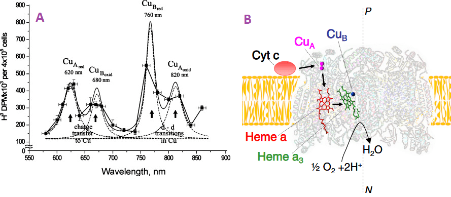
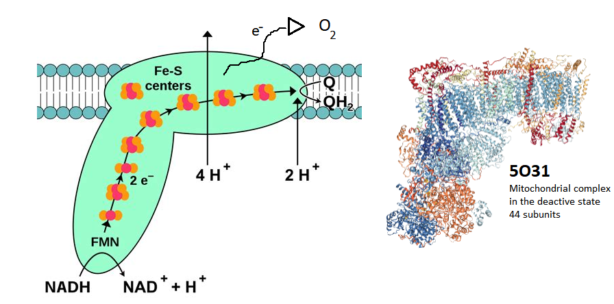
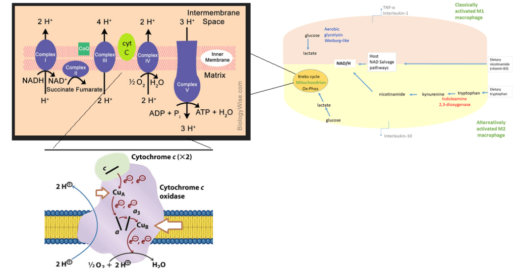
Back in 1997 researchers at the Rocheseter General Hospital irradiated isolated rat liver mitochondria fith 660 nm light from an argon dye laser. [1] They found increased activities of NADH:ubiquinone oxidoreductase (ComplexI), ubiquinol: ferricytochrome C oxidoreductase (Complex III) and ferrocytochrome C: oxygen oxidoreductase (Complex IV) (0.6 J/cm2, 1.2 J/cm2, 2.4 J/cm2 and 4.8 J/cm2, P < 0.05). The activities of succinate ubiquinone oxidoreductase (Complex II), ATPase (Complex V), and lactate dehydrogenase were not affected by photo irradiation. [1] Many compounds that transport electrons also absorb visible light. These include heme groups and Fe-S centers in complexes I-IV. While complex IV has two heme groups, only complex IV has copper. Trying to determine if these compounds absorb at 660 and 820 nm is difficult. Complex IV lives in phospholipid membranes rather than in free solution. Red wavelengths of light tend to scatter more.
Figure 1. The A. The action spectrum for stimulation of DNA synthesis rate on cellular level. Suggested absorbing chromo phores of the photo acceptor, cytochrome c oxidase, are marked from a review of Tiina Daru [1] Note the Y-axis is not dimensional less absorbance units that many scientists are used to seeing. B. Light absorbing cofactors in complex IV with nitric oxide bound [2].
Figure 1A [2] shows what appears to be an absorption spectrum. It is not. The authors are measuring H3 incorporation into DNA in cells grown in heavy water. H32O. Dr Karu has gone into greater detail as what these “action spectra” are all about. This online publication tells us that Dr Karu made many of these assignments prior to the X-ray structure of cytochrome C oxidase.
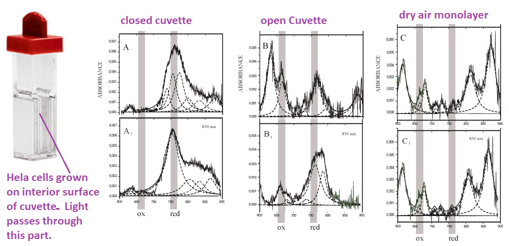
Figure 2, Adapted from a Karu online publication. Note the change in absorbance after irradiation with 830nm changes the absorption spectra. The noisy lines are the actual absorbance spectra. The smooth, dashed lines are curve fitting estimations.
820 nm band belongs mainly to oxidized CuA, prior work by Karu, Cu2+-formate, a surrogate for Cu2+ interacting with a carboxyl group, absorbs in this region.
the 760 nm band to reduced CuB, the
680 nm band to oxidized CuB
620 band to reduced CuA, free Cu2+-(his)2 complexes have absorption peaks at 645nm.
400-450 nm band is more likely to be the envelope of a few absorption bands in the range 350-500 nm (i.e., a superposition of several bands).
404-420 nm oxidized heme,
450 nm region, reduced CuB.
Some experiments…
This red light therapy has been FDA approved for wrinkles, wound healing, and hair growth. Would a MitoCopper cream enhance the red light therapy? Would it kick up the mitochondria in those hair follicles into high gear or simply supply copper to lysyl oxidase?These experiments will not answer the big questions but will hopefully tell you whether or not the MitoCopper cream is working for you. If you truly wanted to be scientific, you’d use four treatments: (1) no treatment, (2) just red light, (3) just MitoCopper cream, (4) MitoCopper cream with red light.
Assuming that you do not want to draw squares on your hair thinned scalp with a black Sharpie, find landmarks on your scalp to guide in the application of your copper cream. Leave a similar sized region as a control. Use your red light as per the instructions. Take photos each day before applying the copper cream and using the red light.
If you are using red light therapy to dissipate post pregnancy stretch marks, Pick a particular set of marks to apply CopperOne cream. Take photographs before applying the cream and using the red light.
References
Yu W, Naim JO, McGowan M, Ippolito K, Lanzafame RJ. (1997) Photomodulation of oxidative metabolism and electron chain enzymes in rat liver mitochondria. Photochem Photobiol. 1997 Dec;66(6):866-71
Tiina I Karu TI (2010) Multiple roles of cytochrome c oxidase in mammalian cells under action of red and IR-A radiation. IUBMB Life 2010 Aug;62(8):607-10. free article
Sarti P, Forte E, Mastronicola D, Giuffrè A, Arese M. Cytochrome c oxidase and nitric oxide in action: molecular mechanisms and pathophysiological implications. Biochim Biophys Acta. 2012 Apr;1817(4):610-9. PMC free article
Bhattacharya, M., & Dutta, A. (2019). Computational Modeling of the Photon Transport, Tissue Heating, and Cytochrome C Oxidase Absorption during Transcranial Near-Infrared Stimulation. Brain sciences, 9(8), 179. PMC free article
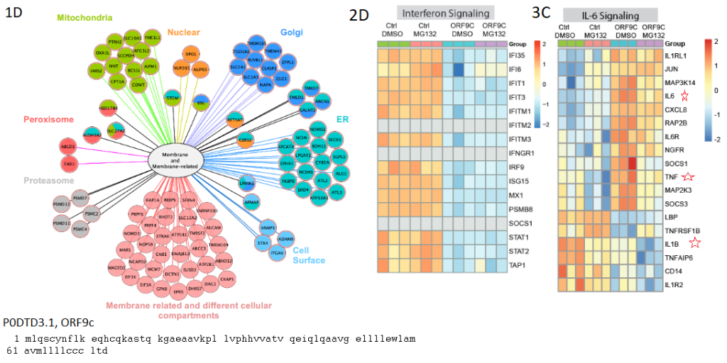
Browsing a January 2021 review on endothelial dysfunction and a potential role of chronic oxidative stress [1] that echoed much of what we’ve had to say about Cu/Zn turning on and off inflammasomes that produce IL1β. They discussed loss of Covid-19 receptor ACE2,more angiotensin II and more super oxide by way of NADPH oxidase. We addressed SOD3 vs the renin angiotensin system in a a slightly different context. These authors led us to ways of looking at Covid-19 proteins. They cited references of papers demonstrating interactions between Covid-19 proteins ORF9c and NSP7 and mitochondrial complex 1 protein NDUFB9 and assembling assisting proteins NDUFAF 1 & 2 that are proposed to be a source of oxygen radicals. [1].
Complex 1 contains at lest 44 protein subunits, one of which is the protein product of the NDUFB9 gene. This gene product is also known as NADH dehydrogenase [ubiquinone] 1 alpha subcomplex subunit 3, the 38 of 46 units. Electrons start at the bottom of the complex and migrate their way to the end to reduce Coenzyme Q. If Covid protein is binding to this complex1 protein, could electrons be diversted to O2?
Some of this is very speculative. If Covid-19 proteins ORF9c and NSP7 bind to Complex 1 proteins in the cell, do they prevent the assembly of complex 1, divert electrons to O2, or nothing at all? Follow this link to learn more about Complex1. Let’s take a look at the molecular fishing expedition.
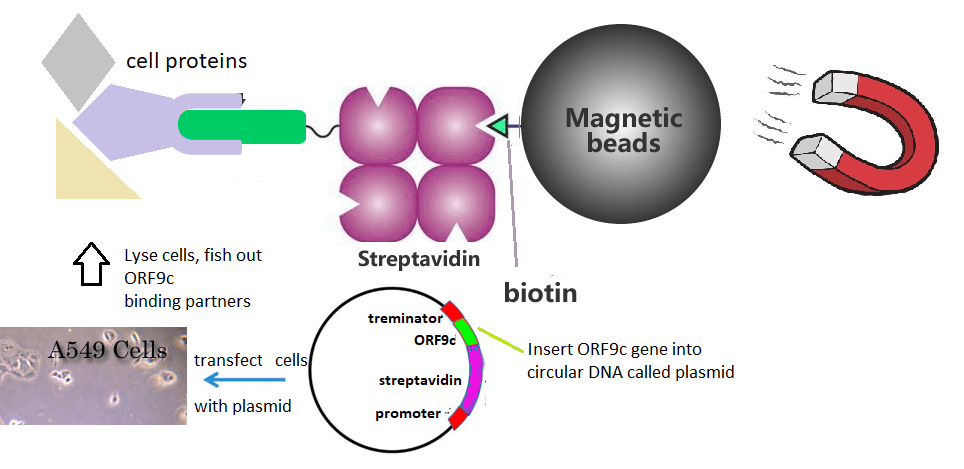
The fishing expedition explained…
The ORF9c protein with an N-terminal biotin tag was expressed in A549 lung cancer epithelial cells. [1] Streptavidin has a very high affinity for biotin. was used to fish out the ORF9c and what ever mammalian proteins might be binding to it. The authors found that ORF9c binds to many membrane proteins in multiple cell organelles. [2] These authors also found that ORF9c decreased stress and immune related transcripts. [2]. It may be too soon to say if Long Covid is a low grade chronic infection.
The search for binding partners of the protein product of the Covid-19 ORF9c gene in extremely simple terms…
Dominguez and coworkers quickly discovered that the ORF9c gene product associated with proteins in membranes of sub cellular organelles [2]. The authors addressed homology with other membrane associated proteins [2]. They also addressed how expressing this Covid-19 protein in lung epithelial cells, with or without the proeosome inhibitor MG132, affected gene transcription as well as proteins. [2] MG132 was used to inhibit protein degradation via the unfolded protein response. Below are some exerts from a very detailed paper that came to the conclusion that the protein product of the ORF9c gene may help Covid-19 evade the immune system. [2]
Netting cytokine and immune system evasion fish
1D membrane proteins found associated with the ORF9c/svidin chimera from Fig 1D of ref [2] 2D Translated proteins that increased or decreased in abundance. 3C chages in mRNA transcript in IL6 pathways. [2]
Note that the control was a chimera of avidin and a small protein called green fluorescent protein. This serves as a nice control for epithelial cells responding to the translation of unnatural proteins. Changes in protein levels may be due to changes in the translation of mRNAs or to changes in degradation, hence the proteosome inhibitor MG132. STAT1 and STAT2 are are transcription factors that control the expression of interleukins among other proteins. The interferon reducible (IGI) family of proteins will not be addressed in this post. They are involved in Note the increase of cytokine transcripts as well as their receptors in GFP-avidin transfected cells versus ORF9c-avidin transfected cells. Note that the TAP1 protein is down regulated with or without proteosome inhibition. The Herpes virus evades the immune system by blocking the TAP1 protein from transporting viral peptides to the cell surface to alert circulating T cells. Down regulating this protein may have the same effect as blocking it.
References
Chang, R., Mamun, A., Dominic, A., & Le, N. T. (2021). SARS-CoV-2 Mediated Endothelial Dysfunction: The Potential Role of Chronic Oxidative Stress. Frontiers in physiology, 11, 605908. Link
Dominguez Andres, A., Feng, Y., Campos, A. R., Yin, J., Yang, C. C., James, B., Murad, R., Kim, H., Deshpande, A. J., Gordon, D. E., Krogan, N., Pippa, R., & Ronai, Z. A. (2020). SARS-CoV-2 ORF9c Is a Membrane-Associated Protein that Suppresses Antiviral Responses in Cells. bioRxiv : the preprint server for biology, 2020.08.18.256776. Cross Ref
Encephalitis is the inflammation of the brain usually brought on by a viral infection. The N-methyl-D-aspartate receptor is an ion channel for Na+ and Ca2+ that is gated by the synthetic ligand NMDA. Its natural ligands are glycine/D-serine and glutamate. Influx of Na+ and Ca2+ into the cell causes the neuron to depolarize sending an action potential down its axon that results in the release neurotransmitter into the synapse in the vicinity of the dendrites of the post synaptic neuron. Opening and closing of the pore that allows the passage of Na+ and Ca2+ into the cell is gated by the binding of glutamate (Glu) and glycine.
Figure 1 The NMDA receptor A. The NMDA receptor consists of two subunits: NR1 and NR2. Auto antibodies are against NR1 The 5 potential glycosylations were not in the original reference. B. Domains of the NR1 receptor form UniProt.org. C. The typical IgG auto antibody is anticipated to be 50% larger than the NR1 subunit of the NMDA receptor
The authors of a recent review on NMDA receptor encephalitis discuss the correlation of the symptoms with the Ab titers in the CSF rather than the serum. [1]. Teratomas contain multiple cell types and may arise from the ovary or testicle.
A tumor may express the neuronal ion channel. Ovarian teratomas were given as an example. [1]
Herpes simplex virus infections were given as another example. [1] The authors did not speculate as to whether or not a virus must infect neurons in order to trigger production of auto antibodies against neuronal surface proteins.
Unlike the example of the streptococcal M protein resembling cardiac myosin components in rheumatic heart disease, we don’t have an example of compoents of any viral protein resembling the NR1 subunit of the NMDA receptor.
Genetic susceptibility was mentioned as a cause. [1]
Auto antibody binding to the NMDA receptor was mentioned as resembling pharmacology. [1] Which pharmaceuticals? Let’s look at NMDA receptor antagonists. We have the competitive inhibitor Aspartame, the artificial sweetener with neurological side effects. Gabapentin, the anti-convulsant, is a noncompetitive inhibitor. Among NMDA receptor agonists we have the allosteric modulator cholesterol and the partial agonist NMDA.
A journey of the imagination
Just looking at the cartoon in Figure 1A we see that extracellular domain S2 has six potenital glycosylation sites that may offer steric protection from auto antibody binding. Antibodies against the N-terminal domain (NTD) could conceivably block access of glycine. .. or mimic the binding of glycine. Receptor cross linking with a single IgG, what would be the consequences? Some might say receptor endocytosis. The physician treating a patient with unexplained symptoms might order an auto antibody panel to test the CSF. Many of these amateurish questions have been asked in a more sophisticated manner by Dr. Yue-Qiao Huang of Philadelphia College of Osteopathic Medicine Georgia School of Pharmacy and coauthors and coauthors. [2] This review is truly comprehensive and covers many animal models. [2] How do these auto antibodies get past the blood brain barrier in the first place?
Getting auto antibodies past the blood brain barrier
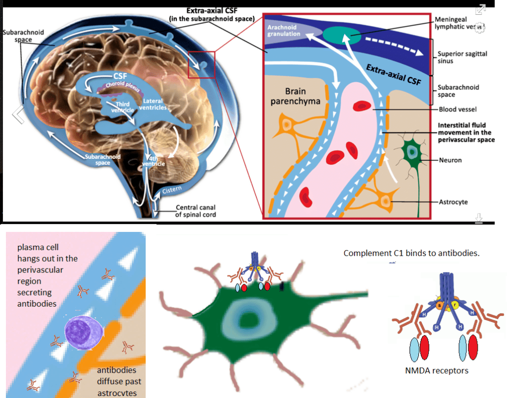
The Wandinger review briefly mentioned a small study that partially answered this question. [1] This study looked for antibody producing plasma cells in the brains of deceased patients who had auto immune encephalitis [3] Some physicians will learn that the blood brain barrier is far more complicated than they may have been taught in medical school. The two main findings of this study were
Syndecan / CD138+ cells were found in the brains of diseased patients with anti-NMDAR encephalitis. Syndecan is an extracellular membrane attached protein that was used as a marker for antibody producing plasma cells.[3]. While syndecan may be expressed in multiple organs and cell types, its immuno reactivity was was not found in control brains in this study. [3] These authors reported syndecan staining cells in Virchow-Robin spaces. [3]
These authors also reported complement staining in the brains of of diseased patients with anti-NMDAR encephalitis. [3].
Figure 2, Interpretation of reference [2]. The perivascular space image is from Wikipedia. Antibody secreting plasma cells were shown to hang out in this space, presumably attached to walls using syndecan/CD138. Secreted antibodies can presumably diffuse past the astrocyte barrier to bind to the NR1 subunits of NMDA receptors on neurons. Complement binding was detected in this region.C1 is fllowed by C2-4 and eventually the membrane attack complex C5-9.
Does auto antibody binding to the NMDA receptor mimic pharmaceuticals and/or initiate a membrane attack complex? Most standard treatments seek to turn off all immune response or counter all antibodies, auto or against foreign antigens. Is there a diet that mitigates the migration of plasma cells into this perivascular space?
Case studies
In many of these reports there is a lack of certainty of a causal relationship between a Covid-19 infection and anti-NMDA receptor antibodies. Perhaps the Coivd-19 infection triggered the movement of plasma cells programmed to produce anti-NMDA receptor antibodies into the perivascular spaces of the brain. Did the Covid-19 infection trigger the production of these auto-antibodies?
Case reports of Covid1=9 concombinant with anti-NMDA receptor encephalitis
Management
Management of autoimmune encephalitis is pretty much as we have discussed so far. [10]. This review discusses the pharmacology of antibodies binding to various neurotransmitter receptors that goes beyond what we have discussed so far. [10], Autoimmune-enc ephalitis.org has some interesting things, both positive and negative, about the keto diet. Aside from the ketogenic diet, there really isn’t a diet for autoimmune encephalitis.
References
Wandinger KP, Leypoldt F, Junker R. (2018) Autoantibody-Mediated Encephalitis.Dtsch Arztebl Int. 2018 Nov 5;115(40):666-673. PMC free article
Huang, Y. Q., & Xiong, H. (2021). Anti-NMDA receptor encephalitis: a review of mechanistic studies. International journal of physiology, pathophysiology and pharmacology, 13(1), 1–11. PMC free article
Martinez-Hernandez, E., Horvath, J., Shiloh-Malawsky, Y., Sangha, N., Martinez-Lage, M., & Dalmau, J. (2011). Analysis of complement and plasma cells in the brain of patients with anti-NMDAR encephalitis. Neurology, 77(6), 589–593. PMC free article
Moideen, S., Thomas, R., Suresh Kumar, P. N., Uvais, N. A., & Ul Haq Katshu, M. Z. (2020). Psychosis in a patient with anti-NMDA-receptor antibodies experiencing significant stress related to COVID-19. Brain, behavior, & immunity – health, 7, 100125 PMC free article
Panariello, Adelaide et al. “Anti-NMDA receptor encephalitis in a psychiatric Covid-19 patient: A case report.†Brain, behavior, and immunity vol. 87 (2020): 179-181. PMC free article
Monti, G., Giovannini, G., Marudi, A., Bedin, R., Melegari, A., Simone, A. M., Santangelo, M., Pignatti, A., Bertellini, E., Trenti, T., & Meletti, S. (2020). Anti-NMDA receptor encephalitis presenting as new onset refractory status epilepticus in COVID-19. Seizure, 81, 18–20. PMC free article
Burr, Tyler et al. “N-Methyl-d-Aspartate Receptor Encephalitis Associated With COVID-19 Infection in a Toddler.†Pediatric neurology vol. 114 (2021): 75-76. PMC free article
Sarigecili, E., Arslan, I., Ucar, H. K., & Celik, U. (2021). Pediatric anti-NMDA receptor encephalitis associated with COVID-19. Child’s nervous system : ChNS : official journal of the International Society for Pediatric Neurosurgery, 1–4. Advance online publication. PMC free articles
McHattie, A. W., Coebergh, J., Khan, F., & Morgante, F. (2021). Palilalia as a prominent feature of anti-NMDA receptor encephalitis in a woman with COVID-19. Journal of neurology, 1–3. PMC free article
Macher S (2018) Management of Autoimmune Encephalitis: An Observational Monocentric Study of 38 Patients. Front. Immunol., 22 November 2018 PMC free article
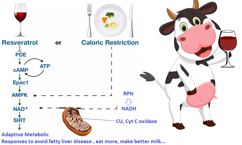
Some of the information in the post may be new to the average dairy farmer. Our featured image, adapted from sirtuins for human health, is a good introduction to what this post is about. The dairy farmer may have heard that drinking resveratrol rich red wine is a good way to get the same benefits of caloric restriction as they both activate the longevity enzyme called sirtuin 1, or Sirt1 for short. Why is negative energy balance so bad for cows? Maybe it is not so bad if the cow has the right nutrition. We will argue that the rumen protected niacin and copper supplements you’ve been giving your cows have some interesting science behind them.
When cows restrict their food calories
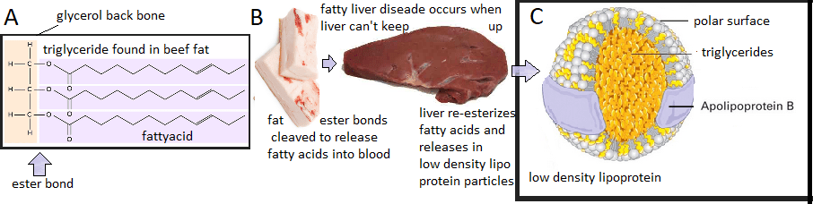
According to the MSD Vet Manual fatty liver disease in cattle occurs during a time of negative energy balance, i.e. when the cow is tapping into triglyceride fat stores to make up for a deficit of food calories. Calving and going off feed are times when a cow can experience a negative energy balance.
Figure 1. Fat shuffling in cows. A. Triglycerides are a glycerol backbone (orange) with three fatty acids (purple). B. Images of bovine triglyceride fat stores from which fatty acids are released into the blood stream to be repackaged into C. triglyceride containing low density lipo protein particles that are released into the cow’s blood stream.
When a cow is burning more calories than she consumes, she taps into her fat stores by releasing the fatty acids from triglycerides. The liver takes these fatty acids, puts them back onto glycerol backbones, and then packs them into low density lipoprotein particles to be released into her blood stream. Sometimes the cow’s liver just cannot keep up. Cows eating too much does not cause fatty liver disease, but not eating does according to msdvetmanual.com. While some humans might restrict their calorie intake to tap into fat reserves, this is not a common practice of lactating humans. We will discuss what happens in cows when they are supplemented with rumen protected niacin.
Niacin in fatty liver disease
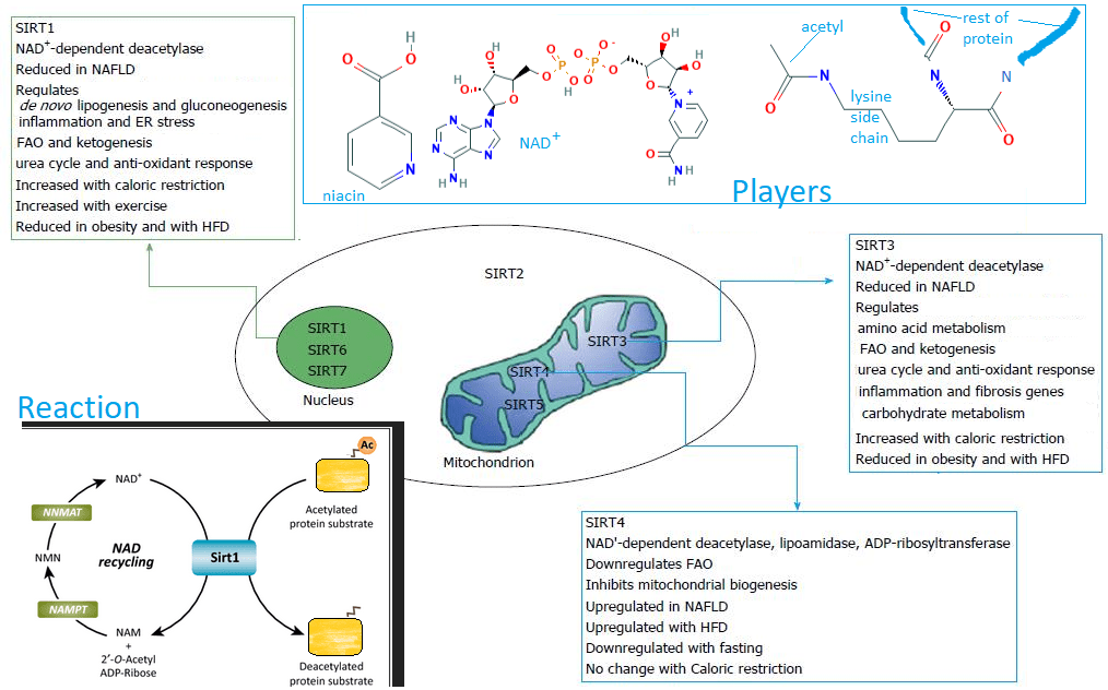
In a previous post we reviewed a study showing the ability of Cu(I)NA2 to mitigate fatty liver disease in a rat model. We neglected the potential niacin contribution to the story. Nassir and Ibadh have a nice review on the role of “longevity” enzymes called sirtuins in human fatty liver disease. [1] We have modified a summary figure from this review to illustrate the deacetylation reaction and the players.
the players
Niacin, vitamin B3, is the other two thirds of Cu(I)NA2. It is also a component of nicotinamide adenine dinucleotide NADH/NAD+. We have covered reduction of TCA cycle generated NADH to NAD+ in a post in a introducing sirtuins and other NAD+ dependent enzymes.
Figure 2 Sirtuins in human fatty liver disease adapted from reference [1] . We have added the following images. Players niacin a precursor of NAD+, and the lacerated lysine side chain found in proteins . The reaction Sirtuins, of which Sirt1 is one, remove acetyl groups (Ac) from nitrogen of lysine side chains. High fat diet, HFD; non alcoholic fatty liver disease, NAFLD; fatty acid oxidation, FAO; endoplasmic reticulum, ER;
The reaction, for the sake of our discussion, is simply the removal of acetyl groups from lysine side chains of proteins. These proteins may be histones, the spools on which chromatin DNA is wound. The acetyl group abrogates the positive charge of the of the lysine group. Make careful note of the role of NAD+ dependent sirtuins in shuttling fats in and out of the liver but also activation of genes involved in anti-oxidant response. [1] Make careful note that human Sirt1 is reduced by high fat diets (HFD) and increased by exercise and caloric restriction. [1] Some Chinese investigators asked,
“What happens to Sirt1 in peripartum dairy cows with mild fatty liver?”
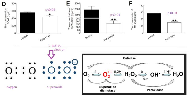
Dairy cows were classified based on their hepatic triglyceride content. The fatty liver cows’ triglyceride content was about 2% of the liver weight whereas the control cow’s were slightly under 1%. [2] Let’s simplify this discussion of a very complicated paper by looking at a few panels of Figure 3 at a time. These data are enzyme activities.We are about to learn that enzyme activity of anti oxidant enzymes also takes a hit. From the description in reference [2] liver samples were homogenized. Enzymes from soluble fractions were capture with antibodies in ELISA plates and analyzed for their respective activities.
Enzymes that control gene tanscription
Sirt1 gets placed in this category because histone acetylation influences gene transcription.
Figure3, from referece [2]. Panels A-C of this publicatgion ar te enzymes that influence gene transcription. Note that even when the change is significant, the change is. not that large
peroxisome proliferator-activated receptor γ coactivator-1 alpha (PGC-1α),in simple terms binds fatty acids and helps regulate gene transcription. Sterol regulatory element binding proteins (SREBP) are also transcription factors that bind to the regulatory elements of genes. SREBP gets its start in membranes as bHLH-Zip. When cleaved, bHLH-Zip/SREBP reports back to the nucleus to regulate cholesterol related gene transcription.
Anti-oxidant enzymes
Catalase is an iron containing enzyme that catalyzes the decomposition of hydrogen peroxideH2O2 to water H2O and molecular oxygen, O2. Superoxide dismutases catalyze the dismutation of super oxide to O2 and H2O2. In Cu/Zn SOD the Zn is only structural. Cu2+-SOD + O–2 → Cu+-SOD + O2 followed by Cu+-SOD + O−2 + 2H+ → Cu2+-SOD + H2O2 . Manganese cofactor SOD is found in the mitochondria
Figure 3 cont from reference [2] and embelished with images. These panels tell the story of enzymes that detoxify superoxide, a reactive oxygen species. Note the large decrease in Cu/Zn superoxide dismutase (SOD)
Four electrons participate in a double bound between the two oxygen molecules in molecular oxygen, O2. Super has only a single bond between the two oxygens. An additional electron completes the eight electron valence shell of one oxygen leaving the other oxygen with an unpaired electron. This unpaired electron makes super oxide a reactive oxygen species. Note the remarkable decrease in Cu/Zn SOD and the break in the Y-axis of panel E needed to document the >10x decrease in Cu/Zn SOD activity in bovine fatty liver disease.
Thiol redox balance and reactive oxygen species
Glutathione peroxidases are a family of selenocysteine containing enzymes serve the same function of catalase: conversion of H2O2 to H2O. They also convert lipid peroxides to their corresponding alcohols.
Figure 3cont. from reference [2] plus acon n overview of redox enzymes.
These authors saw a significant, but not so large, decrease in Sirt1 activity in cows with mild fatty liver disease. They did see large decreases in the Sirt protein and mRNA (not shown in this post). These authors saw decreases in the following transcripts in fatty livers:
Mn SOD, ~20x
catalase, ~5x
glutathione peroxidase, ~10x
Cu/Zn SOD, ~ 3x
Unlike Cu/Zn SOD activity, the changes in Cu/Zn SOD transcripts did not reach the threshold of significance at p<0.05. This publication was far more focused on changes in transcripts of genes involved in fatty acid metabolismi. Whether the fatty liver condition interfered with Cu/Zn SOD being loaded with metal cofactor was not part of the objectives. [2] A follow up to this study looked at which proteins were acetylated in fatty liver disease in cattle. [3] These authors identified many mitochondrial enzymes involved in fatty acid metabolism. Electron transport enzymes and anti oxidant enzymes were not prominent or even present in this “acetylome.” [3]
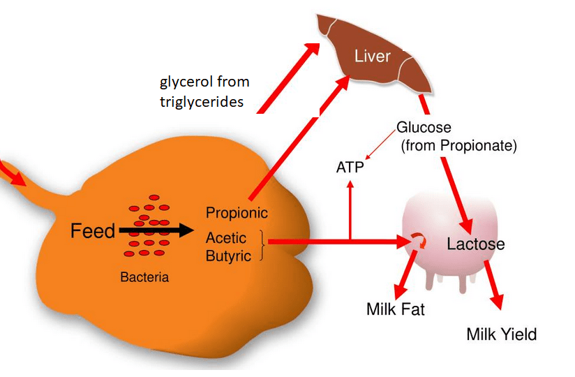
Back to the basics, making milk
Modifying protein function by placing acetate tags on protein lysines is trendy in biological science.
Figure 4 Reminder that cows need to be fermenting feed to make milk. They need glucose to fuel their brains too. Mammalian brains do not break down fatty acids for ATP. They use glucose, and in a pinch, they can use beta-hydroxy butryrate.
This whole discussion of the longevity sirtuin enzymes, them getting enough NAD+ cofactor to deacetylate proteins that somehow get acetylated in the peripartum fatty cow liver seems to be missing the point of making milk! How milk production is responsive to the cow’s energy status will probably continue to keep scientists busy.
RPN increases dry matter intake and milk yield
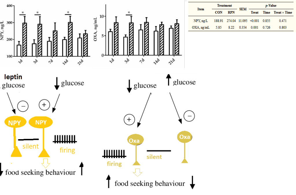
Rumen protected niacin (RPN) has become a popular dietary supplement for milk cows. Chinese dairy farmers observed that cows on RPN simply ate more. [4] Chinese scientists conducted a bovine “clinical” trial of 12 multiparous Holstein dairy cows. The cows were divided into two groups with diet supplemented with either 0 (CON) or 20 g/day RPN (RPN). [4] Each group contained three 3rd parity and three 4th parity cows. Milk production, milk composition, and dry matter intake were the “tried and true” outcome measures in this mini trial. [4] 21st Century outcome measures were appetite stimulating homores neuropeptide Y (NPY), orexin A (OXA), non-esterified fatty acids (NEFA), β-hydroxy butyric acid (BHBA), and rumen bacteria counts.
eating more and making more milk
Figure 5 from reference [4] Graphical and tabular data have been combined.
The neurotransmitter revolt?
How calorie restricted were these cows? When cows are niacin supplemented, peptide Deeurotransmitters that increase appetite are relased. Going back to the Wikipedia links, orexin and neuropeptide Y secreting neurons depolarize in response to lowered blood glucose. Depolarization translates into increased firing and neuro transmitter release. NPY secreting neurons are inhibited by glucose and leptin, a peptide secreted by fat cells.
Figure 6 graphical data from reference [4] showinspg changes in neurotransmitters in response to rumen protected niacin. Images have been added to explain the relationship to glucose.
“Brain food” from fatty acids requires NADH
Non-esterized fatty acids (NEFA) have been covered. Free fatty acids are taken apart two carbons at a time in a process called beta-oxidation. Acetyl-CoA feeds into the TCA cycle that generates NADH to be used by the electron transport chain. Alternatively, two acetyl-CoA may condense to form acetoacylCoA. This compound is further reduced by NADH to form beta-hydroxy butyric acid. BHBA can be used as a fuel for the brain when the cow in a state of negative energy balance.
Figure 7 Data from refereence [4] Non-esterized fatty acids (NEFA) are converted to acetyl CoA via beta-oxidation. Acetyl CoA is a precursor for beta hydroxy butyrate. Data in the yellow table are from ANOVA analysis that examines interactions between time and treatment, niacin versus control.
The authors stated that none of their cows reached the threshold of NEFA and BHBA seen in fatty liver disease. Small increases in NEFA and the corresponding BHBA may be a good thing if it keeps the cow’s brain going until it can produce more NPY and/or orexin to stimulate her appetite. If she eats more, her rumen will produce more propionic acid that her liver will use to make glucose, proper brain food and a precursor for lactose for her milk.
Supplementing cows with copper because our soils are soils are depleted
Dairy farmers can probably teach us more than a few things about this problem. We have addressed our copper depleted soils on the Copper Electron Thesis page. We have visited company websites that sell copper supplements to rancher. Copper is needed for proper flow of electrons through the electron transport chain to ATP. Remember that these cows go through a spat of eating less so there is less propionate to make glucose. Anaerobic glycolysis is not a favorable option. Their mitochondria may be generating more superoxide than usual. Cow’s need Cu/Zn and Mn SOD to clean up the mess.
A milk cow supplement containing copper and niacin?
What are your thoughts on rumen protected copper? Would you be interested in supplementing your cows with rumen protected copper and niacin? Among many other things, copper is needed for proper flow of electrons.
References
Nassir F, Ibdah JA. (2016) Sirtuins and nonalcoholic fatty liver disease. World J Gastroenterol. 2016 Dec 14;22(46):10084-10092 PMC free article
Li Y, Zou S, Ding H, Hao N, Huang Y, Tang J, Cheng J, Feng S, Li J, Wang X, et al. Low expression of sirtuin 1 in the dairy cows with mild fatty liver alters hepatic lipid metabolism. Animals (Basel) 2020;10(4):560. PMC free article
Le-Tian, Z., Cheng-Zhang, H., Xuan, Z., Zhang, Q., Zhen-Gui, Y., Qing-Qing, W., Sheng-Xuan, W., Zhong-Jin, X., Ran-Ran, L., Ting-Jun, L., Zhong-Qu, S., Zhong-Hua, W., & Ke-Rong, S. (2020). Protein acetylation in mitochondria plays critical functions in the pathogenesis of fatty liver disease. BMC genomics, 21(1), 435. PMC free article
Gaowa, N., Zhang, X., Li, H., Wang, Y., Zhang, J., Hao, Y., Cao, Z., & Li, S. (2021). Effects of Rumen-Protected Niacin on Dry Matter Intake, Milk Production, Apparent Total Tract Digestibility, and Faecal Bacterial Community in Multiparous Holstein Dairy Cow during the Postpartum Period. Animals : an open access journal from MDPI, 11(3), 617. PMC free article
This post reviews a few recent reports in the literature concerning niacin, the other two thirds of CopperOne, and Covid. Most of the latest are nice reviews and novel ideas. Then there is a review that forces us to say, “Wow! CopperOne may modulate our immune response to more infections than Covid-19.
Some very nice reviews,clever in silico, and in lab experiments
Min Su and coauthors (Guangxi Key Laboratory of Tumor Immunology and Microenvironmental Regulation, Guilin, China) published some interesting ideas concerning the gene overlap of treating colorectal cancer (CRC) and Covid-19 with niacin. [1] We’ve covered a lot of similar concepts in niacin benefits infections. These authors suggested that niacin might actually bind to the inflammatory cytokine IL-1β They came to this conclusion using in silico molecular docking techniques. [1]
The angiotensin converting enzyme 2 is not just for proteolysis of angiotensin II. Camargo and coauthors remind us that this protease can also form heterodimers with the broad neutral ammino acid transporter (B0AT). [2] We are also reminded that mutations in B0AT cause Hartnup Disease which has pellegra like symptoms. If Covid-19 abinds to ACE2 receptors in the small intestine brush borders, the absorption of amino acid precursors of niacin and serotonin might be compromised. [2] Niacin supplementation might be required. [2]
Doroftei and coauthors published an extensive review of niacin/NADH on many biological pathways as they pertain to oxidative stress and Covid-19 infections. [3]
Niacin has been shown to bind to the main 3CLpro protease of Covid-19 and modestly inhibit it’s activity. [4]
And this is how CopperOne works!
Or so we like to think..
This review was authored by Melinda Suchard Dana M. Savulescu of National Institute for Communicable Diseases, Johannesburg, South Africa. Dr Suchard is an expert in the role of indoleamine 2,3-dioxygenase (IDO) in infectious diseases. These authors define “sepsis” as the body’s response to an infection. Septicemia is defined as infectious particles in the blood.
Nicotinamide and macrophage phenotypes
The reducing eqquivalent nicotinamide may be derived from dietary niacin, recycled through existing the reduced version, or synthesized de novo from the amino acid tryptophan. The rateâ€limiting enzyme for de novo nicotinamide synthesis is IDO, a haemâ€containing intracellular enzyme found predominantly in cells of the macrophage/monocyte lineage. [5]
M1 macrophages secrete proâ€inflammatory cytokines such as TNF alpha and interleukin 1â€Î².
M2 macrophages secrete cytokines such as interleukinâ€10, which have immune suppressive functions and play a role in wound healing.
Immunometabolism in macrophages
Resting macrophage utilize the TCA cycle to generate NADH that feeds into the electron transport chain to generate ATP. When stimulated with pathogens macrophage switch to glycolysis. This Warburg metabolism is also found in cancer cells. While not as efficient as aerobic respiration, stopping at pyruvate allows the macrophage to conserve pyruvate for synthesis of other biological materials.
Right images of the role of copper in the mitochondria. Left from ref [5]
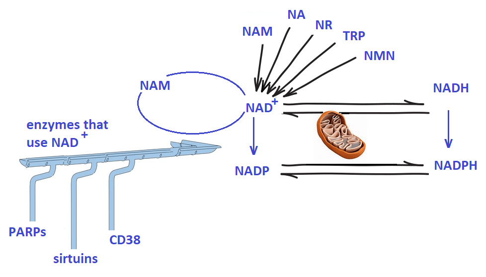
A closer look at pathways intersecting with NAD+
The authors looked at pathways regulated by NAD+, the oxidized form of NADH + H+,. from the standpoint of IDO kicking in, or not, when dietary niacin is insufficient. These pathways include:
PARPADP ribose polymerase enzymes use NAD+ as a substrate to repair DNA double strand breaks.
NAD+ also plays a key role in autophagy through partnering with sirtuins, which are NADâ€dependent deacetylases.
CD38can act as an extracellular receptor for NAD+ and an enzyme that produces ADP ribose from NAD+.
NAD+ is released from cells during early inflammation where it may interact with CD38 on T and B lymphocytes. [5] CD38 may also be found on airway smooth muscle cells. [5]
M1 macrophage activation occurred simultaneously with NAD+ depletion .
Nuclear NAD+ may affect transcription of key inflammatory genes.
Looking forward
The Suchardand Savulescu review on NAD+ pathways in macrophage may change how we view a lot of things beyond Covid and the immune system.
Note the role of both NAD+ and ATPSummary of NAD+ pathways NAM, nicotinamidr; NA, nicotinic acid; NR, nicotinamide riboside; Trp, tryptophan; NMN, Nicotinamide mononucleotide; NAD+, oxidized Nicotinamide adenine dinucleotide; NADH, reduced NAD+; NADP, Nicotinamide adenine dinucleotide phosphate; NADPH, reduced NADP. All NAD+ signalling is very responsive to an active electron transport chain in the mitodhondria. NAD+ may be recycled or siphoned off to other enzymes.
The added component of CD38 signalling, that we are only now becoming aware of, adds a whole new component of how Cu+ and nicotinic acid may act in synergy as an immune modulator agent. And a lot more!
References
Li, R., Li, Y., Liang, X., Yang, L., Su, M., & Lai, K. P. (2021). Network Pharmacology and bioinformatics analyses identify intersection genes of niacin and COVID-19 as potential therapeutic targets. Briefings in bioinformatics, 22(2), 1279–1290. PMC free article
Camargo SMR, Vuille-Dit-Bille RN, Meier CF, Verrey F. (2020) ACE2 and gut amino acid transport. Clin Sci (Lond). 2020 Nov 13;134(21):2823-2833.
Doroftei, B., Ilie, O. D., Cojocariu, R. O., Ciobica, A., Maftei, R., Grab, D., Anton, E., McKenna, J., Dhunna, N., & Simionescu, G. (2020). Minireview Exploring the Biological Cycle of Vitamin B3 and Its Influence on Oxidative Stress: Further Molecular and Clinical Aspects. Molecules (Basel, Switzerland), 25(15), 3323. PMC free article
Gao, J., Zhang, L., Liu, X., Li, F., Ma, R., Zhu, Z., Zhang, J., Wu, J., Shi, Y., Pan, Y., Ge, Y., & Ruan, K. (2020). Repurposing Low-Molecular-Weight Drugs against the Main Protease of Severe Acute Respiratory Syndrome Coronavirus 2. The journal of physical chemistry letters, 11(17), 7267–7272. Free PMC article
Suchard MS, Savulescu DM. Nicotinamide pathways as the root cause of sepsis – an evolutionary perspective on macrophage energetic shifts. FEBS J. 2021 Mar 8. Free article
Part of the challenge of finding a treatment is finding a good diagnosis of the problem. What is the Long Covid problem anyway? Long Covid seems a lot like a continuation of the active infection minus the virus shedding. This post examines some of the basic toxicology outcome measures that may or may not lead us to an understanding and ultimately a treatment.
Cytokine profiles
McFarland and coauthors have made a case for how inflammatory cytokines can lead to nociceptor sensitization in dorsal root ganglia.[1] The dorsal root ganglion, as you may recall, is the nodule containing the cell bodies of afferent neurons of the dorsalroot. Afferent neurons contact somatosensory end organs in the skin, deep tissue and viscera.
the Dx
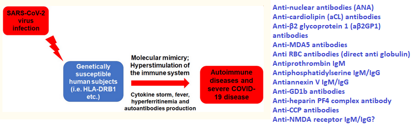
Clinical trials of treatments of active Covid infections have proposed monitoring IL-1β, IL-10, IL-6, IL-18. IL-1β , IL-6, tumor necrosis factor (TNF), and other cytokines were listed as drivers of Long Covid chronic pain. [1] IL-10 is generally considered anti-inflammatory, but has been argued to be pro-inflammatory in Covid-19, cancer, and autoimmune diseases. [2] If the damage is done, what is the point of measuring cytokines as an outcome measure? Long Covid has been proposed to be an autoimmune disease. [3]
Figure 1 adapted from reference [3].
Reference [3] did not mention anti-NMDA receptor antibodies in Covid patients but we have covered thie phenomenon elsewhere on this website. When it comes to chronic pain, it is all about PGE2 that comes after inflammatory cytokines. One question not answered by this review is if auto antibodies binding to their targets sets off a new cytokine storm of diagnostic value.
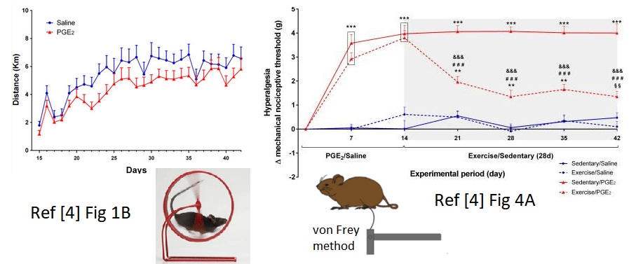
The Rx
In an animal model mouse pindpaws were intraplantar injected with PGE2 or saline in control for 14 consecutive days [4]. After that mice had free access to running wheels for 28 days or were forced to remain sedentary. [4] During the expercise period mechanical hyperalgesia was measured by the electronic von Frey method.[4]
Figure 1, highlights from ref [4] Figure 1B, how much the mice ran on their wheels during the course of he study Figure 4A analgesia induced by voluntary running Data are showed as mean ± SEM (N = 8–10 per group) (***p≤0.001, different from both sedentary/saline and exercise/saline groups; ###p≤0.001, different from sedentary/PGE2 group; **p≤0.01, different from both sedentary/saline and exercise/saline groups; §§p≤0.01, different from exercise/saline group; &&&p ≤ 0.001, different from the time point 14d of the same group)
The images shown n Figure 2 are only a small sampling of a remarkable study. [4] The authors did not attempt to establish a molecular mechanism of exercise induced analgesia. They discussed anti-inflammatory cytokines and the endocannabinoid system as possibilities. Likewise, we do not know if PGE2 DRG sensitization or NMDA auto antibodies are the cause of hyperalgesia. Whether Long Covid hyperalgesia is the result of PGE2 induced modifications of the DRG or auto antibodies, exercise therapy if human patients can be convinced to walk or run 6 km per day like the mice.
Complete blood counts and more
While Long Covid is hitting the peer reviewed literature, there really hasn’t been the extensive work linking parameters uncovered by the complete blood count to symptoms. One study that we know about has followed CBC parameters up to 21 days after symptom onset. [5] At present we are not aware if neutrophil counts stay high and lymphocyte counts stay low in those with the Long Covid syndrome. One would hope that as new treatments for Long Covid undergo clinical trials, CBC will be part of the toxicology tests of an agent on a new population. A complete blood count and all the associated blood chemistry has been conducted on a 57 year old man who presented with autoimmune hepatitis. [5]
We may be required to obtain complete blood counts when testing Cu(I)NA2 on a new population. If we suspect an autoimmune disease, we may want think about sub typing lymphocytes.
Liver Enzymes
The same study reported liver enzymes in the serum of the 57 year old male Long Covid patient.[6]
parameter
patient
reference range
Alkaline phosphatase
48 U/l, repeat 29 U/l
39–117 U/l
Aspartate aminotransferase
137 U/l, repeat 371 U/l
0–40 U/l
Alanine aminotransferase
106 U/l, repeat 246 U/l
0–44 U/l
GGT
655 U/l
0–65 U/l
Prothrombin time
10.4 seconds
9.1–12 seconds
International Norm. Ratio
1
0.8–1.2
Partial thromboplastin time
28 seconds
24–33 seconds
Total iron binding capacity
271 μg/dl
250–450 μg/dl
Iron
222 μg/dl
38–169 μg/dl
Iron saturation
82%
15–55%
Ferritin
860 ng/ml, repeat 3,275 ng/ml
30–400 ng/ml
Erythrocyte sedimentation rate
66 mm/hr
0–30 mm/hr
Haemoglobin A1c
5.90%
4.8–5.6
Anti-smooth muscle Ab
83 units
Positive >30 units
Anti-mitochondrial Ab
174.5 units
Positive >24.9 units
Anti-double-stranded DNA Ab
14 IU/ml
Positive >9 IU/ml
Anti-soluble liver antigen Ab
4.4 units
Negative 0–20 units
Exerts from Table 1 of reference [5] Antibody, Ab; Gamma-glutamyl transferase, GGT;
The authors concluded that this male had auto immune cirrhosis. [7] Liver enzymes are clearly elevated. It is not really clear if the antibodies against smooth muscle proteins contributed to the cirrhosis or if these antibodies affected other organs. Albumin, produced by the liver, was within the reference rage (not shown). Immunoglobins were elevated. Future studies with Cu(I)NA2 to to treat long Covid should include normal blood work that includes liver enzymes. Indeed studies in humans (5-FU toxicity) and rodents (fatty liver model) have shown that Cu(I)NA2 mitigates liver damage.
Standard blood chemistry may be good enough for diagnosing post Covid liver damage. Post Cu(I)NA2 blood chemistry tests may tell us if we have a treatment.
D-dimer
D-dimer is a degradation product of fibrin, the stuff of blood clots. The presence of D-dimer in the blood is an indication of thrombosis, the formation of a blood clot inside a blood vessel. The first report summarized the clinical assessment of 384 patients reviewed a median of 54 (IQR 47–59) days following hospital discharge with COVID-19. [7] Another study found that Long Covid patients with elevated D-dimer tended to be younger and did not require hospitalization. [8] Did the formation of vascular blood clots prevent deleterious internal bleeding? Was vascular damage in the patients with D-dimers long into recovery also severe enough for vascular smooth muscle proteins to be released into the blood and trigger an auto immune reaction? [6]
Figure 3 The stages of clotting. Smooth muscle proteins may be exposed to antigen presenting cells during the initial injury.
If cirrhosis is brought on by damaged blood vessels in the liver, could copper and other nutrient absorption result from damage to blood vessels in the gut? This relates to copper deficiency and eye health problems being brought on by gastric bypass surgery.
A month after the original posting of this post, there have been no new reports of actin auto antibodies in Long Covid to the best of our knowledge. This is probably not something we want to include as a diagnostic of Long Covid and efficacy of Cu(I)NA2.
Dx
We might ask for information s to whether D-dimer was high during the active covid Infection. This might diagnose that vascular damage probably occurred. We can test serum copper bound to ceruloplasmin or something.
Rx
Lysyl oxidase is needed to cross link collagen in damaged blood vessels.
Post treatment Lyme Disease (PTLD) and Long Covid questionnaires
Our previous experience with the Revised Symptom Impact Questionnaire (SQIR) helped us understand how Cu(I)NA2 might be acting in a population with a diverse set of symptoms of nerve/muscle discomfort. Long Covid and PTLD share overlapping symptoms. Might there be a better or an additional questionnaire for long Covid? A previous study used four questionnaires to assess PTLD
Fatigue Severity Scale (FSS) A nine item fatigue metric has summary scores that range from 9 to 63 with a higher score indicating worse fatigue, and with ≥36 indicating clinically relevant levels of fatigue [10]
Short-Form McGill Pain Questionnaire (SF-MPQ) A 15-item pain metric has summary scores that range from 0 to 45 with a higher score indicating worse pain, and with ≥4 indicating a clinically significant experience of pain [11]
Pittsburgh Sleep Quality Index (PSQI) This test has summary scores ranging from 0 to 21 with a higher score indicating worse sleep quality, and with ≥6 indicating clinically significant poor sleep quality. [12]
The Beck Depression Inventory (BDI) is a 21 item depression questionnaire with summary scores ranging from 0 to 63 with a higher score indicating worse depression, and with ≥14 indicating mild, moderate, or severe depression. [13]
The PTLD study concluded that PTLD patients had a lower quality of life than normal controls. [9] These questionnaires may be useful in evaluating Cu(I)NA2 in the treatment of Long Covid-19 and PTLD. Previous experience has suggested that participants might become impatient with too many questions and not give much thought to the answers. A very old questionnaire from 1977 [14] addresses fatigue, pain, and sleep disturbances. [14] The authors were trying to correlate autonomic nervous system function that included “sweaty palms” and “difficulty breathing” with depression and anxiety. [14] Only 24 questions with scores ranging from “very frequently” to “rare or never” may represent less of a challenge to the participant to thoughtfully answer.
Dx tests to keep or not in active vs Long Covid
Cytokine profiles were all the 2020 rage with active Covid, but may or may not be worth the cost and health risks of dealing with infectious blood samples. ORF9c certainly adds an interesting twist if Covid-19 can evade our immune systems in Long Covid.
If Long Covid is auto immune, CBC is a good and inexpensive test to keep.
If Long Covid is auto immune, liver enzymes are a good and inexpensive test to keep.
D-dimer is sort of iffy. The publication source pointed to anti-smooth muscle auto-antibodies that went along with D-dimer. The developing Covid literature is simply not supporting this one.
Quality of Life Questionnaires. Long Covid and PTLD share overlaps. We could ask direct questions as to CDC’s list of active and long Covid symptoms. There is a short and sweet questionnaire that could be informative in conjunction with direct symptom questions that addresses the hypothesis that Cu(I)NA2 helps autonomic nervous system functions. [13]
References
McFarland AJ, Yousuf MS, Shiers S, Price TJ. (2021) Neurobiology of SARS-CoV-2 interactions with the peripheral nervous system: implications for COVID-19 and pain. Pain Rep. 2021 Jan 7;6(1):e885. Free PMC article.
Lu L, Zhang H, Dauphars DJ, He YW.(2020) A Potential Role of Interleukin 10 in COVID-19 Pathogenesis. Trends Immunol. 2021 Jan;42(1):3-5. doi: 10.1016/j.it.2020.10.012. Epub 2020 Nov 2. PMID: 33214057 Free PMC article. Review.
Halpert G, Shoenfeld Y. (2021) SARS-CoV-2, the autoimmune virus. Autoimmun Rev. 2020 Dec;19(12):102695. Free PMC article.
Sartori, C. R., Pagliusi, M., Jr, Bonet, I., Tambeli, C. H., & Parada, C. A. (2020). Running wheel exercise induces therapeutic and preventive effects on inflammatory stimulus-induced persistent hyperalgesia in mice. PloS one, 15(10), e0240115. https://doi.org/10.1371/journal.pone.0240115 PMC free article
Singh, B., Kaur, P., & Maroules, M. (2021). Autoimmune Hepatitis-Primary Biliary Cholangitis Overlap Syndrome Triggered by COVID-19. European journal of case reports in internal medicine, 8(3), 002264. https://doi.org/10.12890/2021_002264 Free PMC article
Mandal S, Barnett J, Brill SE, Brown JS, Denneny EK, Hare SS, Heightman M, Hillman TE, Jacob J, Jarvis HC, Lipman MCI, Naidu SB, Nair A, Porter JC, Tomlinson GS, Hurst JR; ARC Study Group. ‘Long-COVID’: a cross-sectional study of persisting symptoms, biomarker and imaging abnormalities following hospitalisation for COVID-19. Thorax. 2020 Nov 10:thoraxjnl-2020-215818. doi: 10.1136/thoraxjnl-2020-215818. Epub ahead of print. PMID: 33172844; PMCID: PMC7661378. Free PMC article
Lanini, S., Montaldo, C., Nicastri, E., Vairo, F., Agrati, C., Petrosillo, N., Scognamiglio, P., Antinori, A., Puro, V., Di Caro, A., De Carli, G., Navarra, A., Agresta, A., Cimaglia, C., Palmieri, F., D’Offizi, G., Marchioni, L., Kobinger, G. P., Maeurer, M., Girardi, E., … Ippolito, G. (2020). COVID-19 disease-Temporal analyses of complete blood count parameters over course of illness, and relationship to patient demographics and management outcomes in survivors and non-survivors: A longitudinal descriptive cohort study. PloS one, 15(12), e0244129. https://doi.org/10.1371/journal.pone.0244129 Free PMC article
Townsend L, Fogarty H, Dyer A, Martin-Loeches I, Bannan C, Nadarajan P, Bergin C, O’Farrelly C, Conlon N, Bourke NM, Ward SE, Byrne M, Ryan K, O’Connell N, O’Sullivan JM, Ni Cheallaigh C, O’Donnell JS. Prolonged elevation of D-dimer levels in convalescent COVID-19 patients is independent of the acute phase response. J Thromb Haemost. 2021 Feb 15. doi: 10.1111/jth.15267. Epub ahead of print. PMID: 33587810. Cross Ref
Rebman AW, Bechtold KT, Yang T, et al. . The clinical, symptom, and quality-of-life characterization of a well-defined group of patients with posttreatment Lyme disease syndrome. Front. Med. 2017;4:224 10.3389/fmed.2017.00224 [PMC free article]
Krupp LB, LaRocca NG, Muir-Nash J, Steinberg AD. (1989)The Fatigue Severity Scale. Application to patients with multiple sclerosis and systemic lupus erythematosus. Arch Neurol46(10):1121–3.10.1001 [CrossRef]
Melzack R. (1987)The short-form McGill Pain Questionnaire. Pain 30(2):191–7.10.1016/0304-3959(87)91074-8 [CrossRef]
Buysse DJ, Reynolds CF, III, Monk TH, Berman SR, Kupfer DJ. (1989) The Pittsburgh Sleep Quality Index: a new instrument for psychiatric practice and research. Psychiatry Res 28(2):193–213.10.1016/0165-1781(89)90047-4 [CrossRef]
Beck A, Steer R, Brown G. (1996) Beck Depression Inventory – Second Edition Manual. San Antonio: The Psychological Corporation; . [Google Scholar]
Neziroglul F, Yaryura-Tobias JA (1977) Development of an Autonomic Nervous System Questionnaire: Diagnostic Aid in Measurement of Anxiety, Depression, and Aggression. ORTHOMOLECULAR PSYCHIATRY, VOLUME 6, NUMBER 3, 1977, Pp. 265-271 [free article]
Carrie Burdinski MS, an anatomy and physiology professor at Delta College, has an excellent 2 hour Youtube video on everything you need to know to understand the role of the autonomic nervous system in postural orthostatic tachycardia syndrome (POTS). Ms Burdinski does an excellent job of describing the physiology of non tachycardia symptoms of POTS like “brain fog” and loss of temperature control, both symptoms of Long Covid.
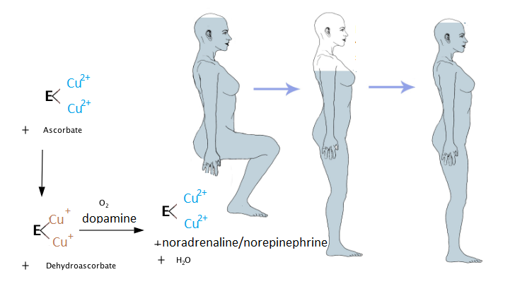
Ms Burdinski gave an interesting overview of different ways that genetics and epigenetics influence the regulation of the norepinephrine (noradrenaline) transporter that clears “used” noradrenaline from the the synaptic cleft. A quick title search of PubMed reveals the following
Mutations in the NET gene that result in a single amino acid substitution in the translated protein that affect the efficiency of noradrenaline / norepinephrine uptake.
Epigenetic methylation of the promoter of the NET gene that prevents translation of the gene into messenger RNA (mRNA)
Epigenetic micro RNA binding to the NET mRNA that prevents it from being translated into a protein.
Once the NET mRNA is translated into a protein, there are numerous “post translational” modifications that can regulate protein function. (Mandela and Ordway 2006)
Examples of regulating NET
These examples from the Mandela and Ordway review are pretty typical of protein regulation. We need to remember that the adrenergic receptors are also subject to regulation at the protein level.
Phosphates may be added to amino acids serine, threonine, and tyrosine by protein kinase C, casein kinase II, Ca2+ calmodulin kinase, and cAMP dependent protein kinase. The latter is particularly interesting because many heterotrimeric G proteins, of which the adrenergic receptors are a few of many, signal through an enzyme that makes cAMP.
A large number of neurotransmitters that bind to heterotrimeric G protein receptors
Protein phosphatases remove phosphates from serine, threonine, and tyrosine.
Insulin is known to decrease NET mRNA expression in locus coeruleus neurons of the rat
Atrial natriuretic factor (ANF) increases production of NET
Nerve growth factor decreases NET mRNA levels.
Nitric oxide may regulate NET by thiol nitrosation and/or cGMP dependent protein kinase.
We will not get into the dozens, if not hundreds, of pharmaceuticals that can regulate norepinephrine/noradrenaline release, reuptake, and binding to its many receptors. Ms Burdinsky was obviously frustrated with the myriad of pharmaceuticals not only to regulate these proteins up to mitigate the side effects.
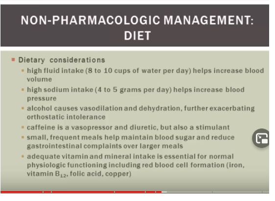
A screen shot of Ms Burdinski’s youtube lecture. This shot covers non pharmaceutical interventions for POTS.
Ms Burdinski went over the physiology behind each and every bullet point on this screen shot and why she thought these dietary interventions help POTS patients. The reasons behind copper were
Copper is needed for proper handling that we have covered in the ceruloplasmin post.
Ms Burdinski postulated that if the presynaptic neuron is that recycling used noradrenaline because of impaired reuptake by NET, it needs more copper for dopamine hydrolase, the enzyme that synthesizes noradrenaline from dopamine. If noradrenaline is not recycled, it just diffuses away from the synaptic cleft. Therefore the neuron needs to make more
Copper deficiency and autonomic dysfunction
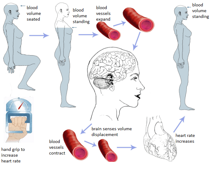
This 1988 study came from the United States Department of Agriculture, Agriculture Human Nutrition Research Center in Grand Forks, North Dakota. Lukaski and coworkers explored this link in female volunteers. This study seemed to start as a simple baroreceptor reflex investigation. When we are at rest, our brains get adequate blood flow. Upon standing gravity decreases the blood volume in our brains. This causes our blood vessels to contract.
When peripheral blood vessels do not properly contract when the woman stands, the brain continues to sense a volume displacement and the heart rate continues to increase. A hand grip exercise was used to increase the heart rate and divert just a small amount of the blood of the women to the exercising muscle.
The diets
Eight women, 18-36 years old, were monitored on four separate diets for a total of 135 days
basal, low diet copper 0.65 mg d -1 and adequate in ascorbic acid (90 mg d-l) 42 days
basal low copper + 1.5 g acid d -1 ascorbic acid for 42 days
basal + 0.8 mg d -1 copper, control normal copper, 14 days
basal +2 mg d –1 copper for 37 days, repletion.
Copper retention or chemical balance
Retention was calculated as the difference between intake and excretion in urine and feces. Menstrual and sweat loses were not considered.
At the end of each dietary period, fasting venous blood samples were obtained to determine biochemical indices of copper status.
Total plasma copper was determined by atomic absorption spectroscopy .
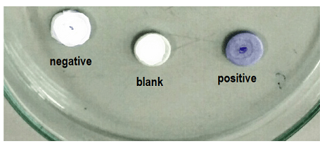
Ceruloplasmin enzymatic activity was assayed as a colorimetric p-phenylenediamine oxidase assay.
Ceruloplasmin content was measured by the radial immuno diffusion assay.
An image of the p-phenylenediamine oxidase assay.An image of the radial immuno diffusion assay. The bigger the circle the greater the activity.
Baroreceptor response
Autonomic cardiovascular function was assessed at the end of each dietary period.
Volunteers were tested in the post absorptive state after a 30-minute rest during which they were supine on a bed in a quiet room.
Variation in resting supine heart rate was determine over a three-minute period using the mean square successive differences of R-R intervals .
Orthostatic responses, upon arising form supine position and standing, were measured for heart rate and blood pressure.
Heart rate response was defined as the ratio of the R-R interval of the 30th to the 15th beat after standing.
Blood pressure was measured as each volunteer was supine and resting quietly and after one minute upon standing.
The hand grip exercise
Standing heart rate and blood pressure responses were determined before and during five minutes of sustained hand grip exercise with the dominant arm at 30% maximal voluntary contraction using a calibrated handgrip dynamometer.
Maximal voluntary contraction was determined at the end of each diet period.
Heart rate was recorded continuously using a multichannel electrocardiograph and standard limb leads.
Blood pressure was determined by auscultation on the inactive arm with diastolic pressure defined as the fourth phase Korotkov sound.
Results
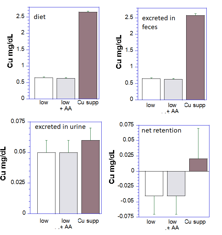
Most copper is excreted in the feces
These data are a graphical representation of Lukaski (1988) table 1. The data from the normal copper arm of the study were not collected, or presented in table 1.
Note that the copper supplemented diet has almost 5x the copper as the low copper diet. A remarkable observation in this study is that whether the copper is low or high, most of it is excreted in the feces. Virtually the same amount of copper is excreted in the urine. Retention of copper is higher in the copper supplemented arm of this study. Ascorbic acid (AA) had no influence on copper retention. What is not clear is if the large excretion of copper is due to failure to absorb it in the first place.
Dietary increases in copper and ceruloplasmin
The following graphs were reproduced from table 2 to emphasize the similarities and differences. Treatments different from the normal copper control at p<0.05 are indicated by “*â€. Variations of copper in these short term diets had no influence on plasma copper. All eight women spent some time in each group.
Data frp, table 2 of the Lukaski publication were graphed to emphasize the changes, and lack thereof
Concentrations of ceruloplasmin, as measured by radial immune diffusion, are in units of mg per liter of plasma. We can only assume that the authors used standards to calibrate the diffusion values. A ceruloplasmin reference range is 200-350 mg per liter. The low copper diet, with or without ascorbic acid (AA) decreased ceruloplasmin activity without decreasing the amount of protein. In fact, AA slightly increased the amount of ceruloplasmin protein in the plasma.
Ceruloplasmin enzymatic activity, in units of mg per liter, dropped (p<0.05) when the participants were on low copper diets with or without ascorbic acid (AA) supplementation. Copper supplementation to 2.65 mg per day did not increase the enzyme activity above what was observed when the participants over on 1.45 mg copper per day (Cu).
POTS spoiler, slight detour
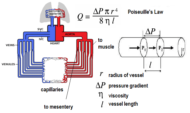
The spoiler alert, the authors reported no differences in the blood pressure responses from going to a supine position to standing. At the time of the study, the scientific community was becoming aware of the cardiovascular response to exercise. A short publication by Qumar and Read (1987) documented a decrease of blood flow to the mesenteric circulation to the small intestine in response to exercise. Many readers were told as children, “Don’t swim right after you eat lunch or you will get a stomach ache!”
If we are running from a dangerous situation, we’d want the blood flow (Q) to our gastrointestinal to decrease somewhat to allow more flow to our legs. When a vessel relaxes, the radius (r) becomes larger and flow increases. When the pressure gradient increases, flow increases. If all vessels were to simultaneously relax, blood flow have to drastically increase to keep the tissues oxygenated. Restricting the flow through some and increasing the flow through those arteries that supply muscles that are exercising means the cardiac output (flow) only has to increase a little bit.
Physical determinants of blood flow, Q. Note that small decreases in the radius of a blood vessel can result in very large decreases in flow. Lines point “to muscle” and “to mesentery.” Imagine another line that points “to brain” for the sake of POTS.
MAP may be estimated as the DP + 1/3 (SP-DP) where SP and DP are the systolic and diastolic blood pressures. It approximates the average pressure during the cardiac cycle. MAP is affected by factors such as:
Volume of blood pumped by the heart per minute (cardiac output, flow, Q)
Heart rate (beats per minute)
Blood pressure
Resistance to blood flow in the vessels
An increase or decrease in any of these factors can proportionately affect mean arterial pressure and bring corresponding consequences to the perfusion of major organs like the brain and kidneys.
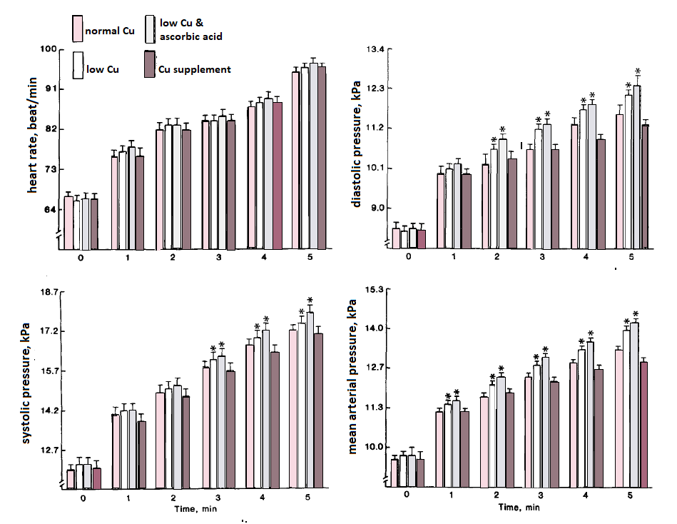
The heart rate was not different before the hand grip exercise started at time 0. The heart rate gradually increased during the 5 minutes of the exercise. The amount of copper made no difference. Overall significance of a copper effect (P<0.001) Maximum voluntary contraction was unaffected by copper.
The heart rate was not different before the hand grip exercise started at time 0. The heart rate gradually increased during the 5 minutes of the exercise. The amount of copper in the diet made no difference.
When the subjects were on low copper diets, their diastolic blood pressures increased more within 2 minutes (P<0.05) of hand grip exercise. (P<0.001) When the subjects were on low copper diets, their systolic blood pressures increased more within 3 minutes (P<0.05) of hand grip exercise.
Mean arterial pressure was noticeably elevated in the copper deficient subjects after only 1 minute of the hand grip exercise. Recall from Poiseuille’s Law that it is the pressure differential, ∆P, and resistance that drives flow.
Lukaski and coworkers discussed the possibility of vascular tone being altered by the impaired collagen cross linking enzyme lysyl oxidase. This group were experts in Cu/Zn superoxide dismutase 3 and angiotenin II activation of super oxide generator NADPH oxidase.
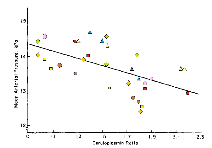
Ceruloplasmin loading of copper predicts MAP in response to exercise
A summary figure related the mean arterial pressure to the hand grip test to the “ceruloplasmin ratio.†This ratio is the enzymatic activity to the amount of the protein as measured by radial immune-diffusion. Each symbol represents an individual participant.
Liberty was taken to draw colored symbols over the poorly resolved black and white symbols in figure 2 of the Lukaski publication. “With the exception of one volunteer in whom this relationship was weak (r=-0.50), the individual relationships had correlation coefficients ranging from – 0.90 to – 0.99 (p<0.01). For the entire study sample, the ceruloplasmin ratio was a significant (p < 0.0004) predictor of mean arterial pressure at the end of the hand grip test.â€
The authors discussed the locus ceruleus as an integrator of afferent input with efferent output that control adrenergic cardiovascular reflexes (locus ceruleus-noradrenergic system or LC-NA system). This locus was also noted as being a copper enriched region of the brain. Blood pressure control during isometric hand grip exercise involves sympathetic (noradrenergic) afferent from skeletal muscle neurons. Lukaski and coworkers (1988) concluded that the involvement of copper required further study.
POTS and Long Covid?
The big question here is if POTS patients and those with Long Covid autonomic dysfunction are copper deficient. David Goldstein of the NIH has reviewed possible causes of POTS in Long Covid. Dr Goldstein gives a more in depth overview of some aspects of the ANS than covered in this post. He mentions autoimmunity. We have covered our research on NMDA repceptor suto antibodies in Long Covid in another post. Blitshteyn and Brook (2017) described a female patient who had received the HPV Cervavix vaccine who later developed POTS. This patient tested positive for anti-NMDA receptor antibodies, responded positively to immunomodulatory therapy, and had her symptoms come back when the therapy was discontinued. The CDC has addressed this association.
Given the prominence of ACE2 in the duodenum and small intestine, could copper absorption via Ctr1 be compromised in some Long Covid patients? Images are from ProteinAtlas.org
POTS and gluten sensitivity?
Copper deficiency in celiac disease has been addressed elsewhere. Are POTS patients more likely to have self reported gluten intolerance or bonefid celiac disease? Hugo Penny and coworkers (2016) of the Royal Hallamshire Hospital in Sheffield, UK surveyed 100 POTS patients. Four of the 100 POTS patients had serologically and biopsy proven celiac disease. Gluten sensitivity was reported in 42% of these 100 POTS patients versus 19% of the control population.
References
Blitshteyn S, Brook J. (2017) Postural tachycardia syndrome (POTS) with anti-NMDA receptor antibodies after human papillomavirus vaccination. Immunol Res. 2017 Feb;65(1):282-284.
Goldstein D. S. (2021). The possible association between COVID-19 and postural tachycardia syndrome. Heart rhythm, 18(4), 508–509.
Hoggard N, Hadjivassiliou M, West JN, Sanders DS. Is there a relationship between gluten sensitivity and postural tachycardia syndrome? Eur J Gastroenterol Hepatol. 2016 Dec;28(12):1383-1387
Lukaski HC, Klevay LM, Milne DB.(1988) Effects of dietary copper on human autonomic cardiovascular function. Eur J Appl Physiol Occup Physiol. 58(1-2):74-80.
Mandela P, Ordway GA. (2006) The norepinephrine transporter and its regulation. J Neurochem. 2006 Apr;97(2):310-33. doi: 10.1111/j.1471-4159.2006.03717.x. Epub 2006 Mar 15. PMID: 16539676 Free article.
Neselioglu S, Ergin M, Erel O. (2017) A New Kinetic, Automated Assay to Determine the Ferroxidase Activity of Ceruloplasmin. Anal Sci.33(12):1339-1344.
Qamar MI, Read AE.(1987)Effects of exercise on mesenteric blood flow in man. Gut. 28(5):583-7.
We have been asked, “Can CopperOne help Amyotrophic lateral sclerosis (ALS) patients given the rumored success of CuATSM clinical trials? This Australian study was accepting patients with familial or sporadic ALS without specifying which proteins (SOD1, TDP-43, or others) are mutated in the familial version when this post was initiated. As of July 25 2023 the results on clincialtials.gov were returned to quality control issues.
Methods: The ALD pilot study was just that. All 12 patient remained on the ALS drug riluzole. Many of the analyses were performed after the patients had died. [1]
CuATSM and riluzole ALS-TDP (n=5) and ALS-SOD1 (n=1)
riluzole only ALS-TDP (n=4) and ALS-SOD1 (n=2)
Results: Our results revealed no significant difference in neuron density or TDP-43 burden in the motor cortex and spinal cord of patients that had received CuATSM compared to patients that had not. In patients that had received CuATSM, p62-immunoreactive astrocytes were observed in the motor cortex and reduced Iba1 density was found in the spinal cord. However, no significant difference in measures of astrocytic activity and SOD1 immuno reactivity was found with CuATSM treatment. [1]
Discussion: CuATSM does not significantly alleviate neuronal pathology or astrogliosis in patients with ALS.
Let’s go back to the original CopperOne customer question, “Can CopperOne help patients with ALS?” We have not clue! We need to first understand what this protein TPD-43 is, why it mis folds, and what may be done to prevent it’s mis folding. We’ll meet a few new copper proteins along the way.
Mutant, misfolded super oxide dismutase 1(SOD1) protein is commonly associated with ALS. An earlier study suggested that sporadic cases were associated with TDP-43 aggregates that tended not to appear in aggregates familial ALS caused by mutations in Cu/Zn SOD1. [2] Misfolded Tar DNA binding protein (TDP-43) is found in many, as well as some familial, cases of ALS. A search of the protein database of crystal structures reveals few structures with single stranded DNA and many aggregates.
The structure of TDP-43
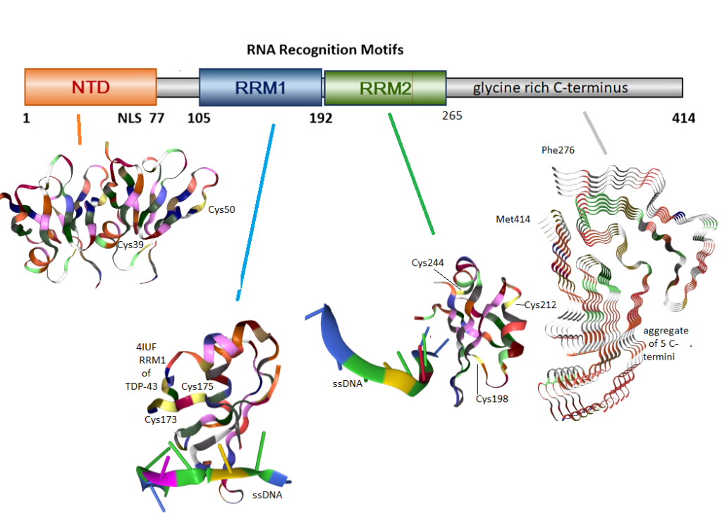
Figure 1 Domains of TDP-43. These images were obtained from rcsb.org. Colors correspond to the type of amino acid. Cysteines are yellow. The N-terminus of TCP-43 has two cysteines which may take the native dimer to a tetramer. The first RNA recognition motif (RRM1) has two cysteines which may form disulfide bonds with each other. RRM2 has three cysteines, two of which have been reported to form aberrant inter-chain cross links. The hydrophobic C-terminus has no disulfides, but can form amyloids as seen in these isolated five fragments.
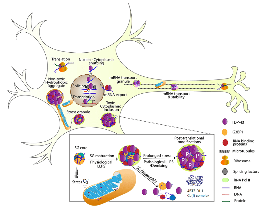
TDP-43 is normally found in the nucleus. A recent review describes its antics in the mitochondria. [2] “TDP-43 proteinopathies†is a term used to describe detergent resistant uniquitinated (figure 2) protein aggregates. As an added bonus, these protein aggregates also tend to be hyper phosphorylated, ubiquitin tags for destruction, and truncated (Figure 1). The accumulation of cytoplasmic inclusion bodies is generally thought of as a toxic gain of function. Formation of these inclusion bodies is accompanied by loss of normal functions that include RNA metabolism,translation of mRNA into protein, stress-induced responses, and mitochondrial function. [3] Studies have demonstrated the association of TDP-43 with mitochondria in motor neuron-like cells. At least a portion of TDP-43 could localize in the inner membrane of mitochondria; full-length and truncated forms of TDPâ€43 could differential reside in the matrix and inter membrane space of mitochondria. [3]
TDP-43 controls mitochondrial trafficking along axons [3], Figure 2
Motor neuron like cells over expressing TDP-43 have reduced mitochondrial complex I activity, mitochondrial transmembrane potential difference, and increased expression of mitochondrial uncoupling protein 2 (UCP2) that functions to decrease the mitochondrial membrane potential.[3]
TDP-43 interacts with mitochondrial RNA
TDP-43 may also facilitate autophagy (self eating) destruction of damaged mitochondria. [2]. The authors mention the Dj-1 protease. As we shall see later, Cu2+ is toxic to proteolytic function.
DJ-1, aka Park7,is a copper cofactor protease localized both in cytoplasm and mitochondria that protects against oxidative stress-induced cell death through the suppression of cytoplasmic TDP-43 aggregation. It was proposed that that the copper enzyme DJ1 may alleviate TDP-43-caused toxicity by degrading both cytoplasmic and mitochondrial TDP-43. [2]
Mitochondrial fission and fusion were discussed in this review [3].
Figure 2 A summary of the many functions of TDP-43. Most of this figure came from [3] with elements of [2] digitally introduced. TFP-43 is mostly a nuclear protein whose interaction with RNA of serves a variety of useful functions [3]. TDP-43 may make its way into the mitochondria where it may disrupt complex 1 as well as disrupt fission and fusion [2]. Ribsomal complexes may also be transported along the axons [3] We suggest that super oxide from dysfunctional mitochondria [2] may release super oxide that is the “stress” trigger for stress granule (SG) core that leads to physiological liquid/liquid phase separation (LLPS).
In a cell culture model Dj-1 co-expression with TDP-43 reduces protein aggregation in response to the mitochondrial toxin and free radical generator paraquat. [4]
TDP-43 Disulfides!
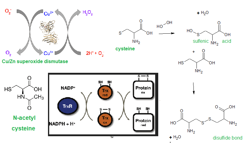
A disulfide is formed when sulfur atoms between two cysteine amino acids form an S-S bond. These disulfide bonds tend to be structural and can be thought of as structural as the buttons and button holes in a dress shirt. Sometimes disulfides form correctly as the new protein comes off the ribosome. In other instances protein disulfide isomerase “unbuttons and rebuttons” disulfides of misfolded proteins in the endoplasmic reticulum. Aberrant disulfides can be formed when sulfur of cysteine is oxidized to cysteine sulfenic acid. This intermediate plays a role in activation of the IL-1β producing inflammasomes.
Figure 3 Cu/Zn SOD 1/3 can be a source of disulfide forming H2O2 as well as detoxifying the much more reactive super oxide. As long as the cell as adequate amounts of catalase, H2O2 will also be detoxified. Thioredoxin (Trx) can use reducing equivalents NADPH + H+ to reduce disulfide bonds.
Let’s take a look at some of the aberrant disulfide bonds of TDP-43 that have been reported in the literature.
Lue71m=,Val72 have been shown to be responsible for non-covalent dimer formation and ability to splice the mRNA for the cystic fibrosis chloride channel CFTR. [5] Two cysteines , Cys39 and Cys50, in this N-terminal domain were shown to be for tetramer formation. [6]
Two forms of oxidative stress, H2O2 and sodium arsenite, have been shown to induce TDP-43 disulfide cross-linking in cultured hippocampus neurons and a cell line expressing TDP-43. Mutants in which Cys198 and Cys244 have been mutated to other amino acids cannot form disulfide bonds. [5]
Cys173 and Cys175 were found to produce intra chain disulfides whereas the others formed inter chain disulfide bonds. [7] Sodium arsentite was used as an indirect means of generating reactive species from the mitochondria.
A mechanism has been proposed by which H2O2 oxidizes one of the two cysteines, Cys198 and Cys244, in the second RNA recognition motif (RRM2). [8]
An extraordinarily through review was published in 2019 by Prasad and coauthors. [9] These authors even covered some publications that suggested TNP-43 aggregation could be protective.
The LOPAC®1280 library, a collection of pharma-developed compounds and approved drugs covering most signaling pathways and major drug target classes was applied at 10 µM concentration to the transfected mouse neuroblastoma Neuro2a (N2a) cells for the TDP-43 self-interaction assay. [10] Auranofin was one compound found to prevent aggregate formation. According to PubChem Auranofin is an inhibitor of thioredoxin reductase, an enzyme thought to give tumor cells resistance to the oxidative stress of their environment.[9] On many levels these results are counter intuitive. Have we not been discussing how inter and intra chain disulfide bond formation can seed aggregation?
The ability of protein disulfide isomerase to prevent aggregation of SOD1 and TDP-43 mutants associated with human ALS adds clarity. [11] Co-expression with functional PDI prevented non-nuclear localization and aggregation in a manner that was dependent on a supply of reduced glutathione.
DJ-1 proteolysis revisited
Protease activity of DJâ€1 lacking Câ€terminal αâ€helix (DJâ€1ΔH9) that is cleaved under oxidative stress conditions was stronger than that of fullâ€sized DJâ€1 [12] The most susceptible sequence digested by DJâ€1ΔH9 was valine–lysine–valine-alanine (VKVA).
Figure 4 from reference [11] DJ-1 is a protease auto-inhibited by a C-terminal helix 9. (top) This study uncovered consensus sites for DJ-1 minus the auto-inhibitory C-terminus (middle). The UniProt sequence of human TDP-43 (bottom) reveals that these DJ-1 sites are found the N-terminus of of TDP-43 that include cysteines involved in tetramer-ization. [12] DJ-1 as three Cu sites. (top) The first Cu site may donate Cu to Cu/ZN SOD1. This site is also essential to proteolysis (middle) Cu may be transferred from the first site to the second (bottom)
The optimal conditions of pH 5.5 and 0 mM NaCl. [12]. Nanomolar Cu2+ was inhibitory to DJâ€1’s protease activity. The authors did not comment on monovalent Cu+. Mutation of the first Cu binding site [13] abolished proteolytic activity[12]. DJ-1 loaded with Cu(II) via CuSO4 was shown to photo oxidize to Cu(I). [13] Transfer between Cu(I) from site 1 to site 2 as well as Cu deficient Cu/Zn SOD1 has also been demonstrated. [13] An unanswered question is if Cu(I), but not Cu(II), can be transferred from site 1 to site 2 so as to enable proteolysis.
A thought experiment summary…
Let’s imagine dress shirts in a wash with all sorts of oxidative stress agents that produce cysteine sulfenic acids that act as button hooks facilitating aberrant disulfide bond (button) formation. The pin stripe dress shirt becomes cross-linked to the paisley, which becomes cross-linked to the plain white shirt, which becomes cross-linked to the plaid. .. To make matters worse, the sleeves of the dress shirts become intertwined and form amyloids like the C-termini of TDP-43 in figure 1. The first response would be to add some thioredoxin to unbutton the mess. Partial unbuttoning would only allow the plaid dress shirt to become cross-linked, button to the plain white dress shirt…Thioredoxin inhibitor Auranofin could be added to the mix to keep the dress shirt aggregate from growing larger. PDI, with its thioredoxin like domains, could be added to our dress shirt wash. PDI unbuttons the aberrant buttoning and allows for the right button-button hole matches. Protease DJ-1 reduces our dress shirt aggregation by ripping some of the dress shirts down the back into two pieces. The role of Cu(I) in this process is kind of murky. Cu(I) in DJ-1 [13] can load Cu/Zn SOD with its Cu cofactor so that the aberrant buttoning does not occur in our wash in the first place.
References
Yang Y, Rowe D, McCann H, Shepherd CE, Kril JJ, Kiernan MC, Halliday GM, Tan RH. Treatment with the copper compound CuATSM has no significant effect on motor neuronal pathology in patients with ALS. Neuropathol Appl Neurobiol. 2023 Jun 15:e12919. PubMed
Mackenzie IR, Bigio EH, Ince PG, Geser F, Neumann M, Cairns NJ, Kwong LK, Forman MS, Ravits J, Stewart H, Eisen A, McClusky L, Kretzschmar HA, Monoranu CM, Highley JR, Kirby J, Siddique T, Shaw PJ, Lee VM, Trojanowski JQ. Pathological TDP-43 distinguishes sporadic amyotrophic lateral sclerosis from amyotrophic lateral sclerosis with SOD1 mutations. Ann Neurol. 2007 May;61(5):427-34. Cross Ref
Gao J, Wang L, Yan T, Perry G, Wang X. (2019) TDP-43 proteinopathy and mitochondrial abnormalities in neurodegeneration. Mol Cell Neurosci. 2019 Oct;100:103396. Free PMC article
Loganathan S, Lehmkuhl EM, Eck RJ, Zarnescu DC.(2020) To Be or Not To Be…Toxic-Is RNA Association With TDP-43 Complexes Deleterious or Protective in Neurodegeneration? Front Mol Biosci. 2020 Jan 10;6:154. Free PMC article.
Lei Y, Zhang ZF, Lei RX, Wang S, Zhuang Y, Liu AC, Wu Y, Chen J, Tang JC, Pan MX, Liu R, Liao WJ, Feng YG, Wan Q, Zheng M. DJ-1 Suppresses Cytoplasmic TDP-43 Aggregation in Oxidative Stress-Induced Cell Injury. J Alzheimers Dis. 2018;66(3):1001-1014
Jiang LL, Xue W, Hong JY, Zhang JT, Li MJ, Yu SN, He JH, Hu HY.(2017)The N-terminal dimerization is required for TDP-43 splicing activity. Sci Rep. 2017 Jul 21;7(1):6196. Free PMC article.
Cohen TJ, Hwang AW, Unger T, Trojanowski JQ, Lee VM. EMBO J. (2012) Redox signalling directly regulates TDP-43 via cysteine oxidation and disulphide cross-linking. Embo J.31(5):1241-52. doi: 10.1038/emboj.2011.471. Epub 2011 Dec 23. PMID: 22193716 Free PMC article.
Rabdano SO, Izmailov SA, Luzik DA, Groves A, Podkorytov IS, Skrynnikov NR.(2017) Onset of disorder and protein aggregation due to oxidation-induced intermolecular disulfide bonds: case study of RRM2 domain from TDP-43. Sci Rep. 2017 Sep 11;7(1):11161. Free PMC article.
Prasad A, Bharathi V, Sivalingam V, Girdhar A, Patel BK.(2019) Molecular Mechanisms of TDP-43 Misfolding and Pathology in Amyotrophic Lateral Sclerosis. Front Mol Neurosci. 2019 Feb 14;12:25. doi: 10.3389/fnmol.2019.00025. eCollection 2019. PMID: 30837838 Free PMC article. Review.
Oberstadt M, Stieler J, Simpong DL, Römuß U, Urban N, Schaefer M, Arendt T, Holzer M.(2018)TDP-43 self-interaction is modulated by redox-active compounds Auranofin, Chelerythrine and Riluzole. Sci Rep. 2018 Feb 2;8(1):2248. Free PMC article.
Parakh S, Shadfar S, Perri ER, Ragagnin AMG, Piattoni CV, FogolÃn MB, Yuan KC, Shahheydari H, Don EK, Thomas CJ, Hong Y, Comini MA, Laird AS, Spencer DM, Atkin JD. (2020) The Redox Activity of Protein Disulfide Isomerase Inhibits ALS Phenotypes in Cellular and Zebrafish Models.Science. 2020 May 22;23(5):101097. Free PMC article.
Mitsugi H, Niki T, Takahashi-Niki K, Tanimura K, Yoshizawa-Kumagaye K, Tsunemi M, Iguchi-Ariga SM, Ariga H.(2013) Identification of the recognition sequence and target proteins for DJ-1 protease. FEBS Lett. 2013 Aug 19;587(16):2493-9. doi: 10.1016/j.febslet.2013.06.032. Epub 2013 Jul 2. PMID: 23831022 Free article.
Girotto S, Cendron L, Bisaglia M, Tessari I, Mammi S, Zanotti G, Bubacco L.(2014) DJ-1 is a copper chaperone acting on SOD1 activation. J Biol Chem. 2014 Apr 11;289(15):10887-10899. Free PMC article.
This post has been updated 04 May 2021. Quoted from the blogpage of NIH director Francis Collins, “If you’d like to find a way to pitch in and help, getting involved in the COVID Symptom Study is as easy as downloading the app.”
How do the symptoms of the active Coivd-19 infection compare with “Long Haul” Covid symptoms that linger after the infection has cleared?
Active Covid-19 versus long haul
Covid-19 active infection [1]
Long haul Covid-19 symptoms [2]
comments
fatigue
fatigue
Shortness of breath or difficulty breathing
Shortness of breath
cough
Cough
Muscle or body aches
Muscle pain
Chest pain
noticeable in active infection?
Joint pain
Rheumatoid arthritis?
Head ache
Head ache
Heart palpitations
Depression
Brain fog
Fever or chills
Intermittent fever
New loss of taste or smell
Smell/taste loss
less common in long haul
Sore throat
Congestion or runny nose
Nausea or vomiting, diarrhea
Comparison of CDC symptoms of active Coivd-19 and long term Covid-19
The gastrointestinal symptoms seem to be restricted to the active infection. Neurological symptoms seem to be features of long haul Covid-19.
According to the CDC less common symptoms may also include cardiac inflammation, renal injury, rashes, and hair loss. Skin rashes and hair loss may also be part of the less common long term symptoms of Covid-19. [2]
Covid-19 and rheumatoid arthritis cytokines
A report from June of 2020 points out that many of the cytokines observed in active Coivd-19 infections are also observed in Rheumatoid arthritis, an autoimmune disorder affecting the joints.[3] According to these authors back in June of 2020, a small percentage of Covid-19 patients experience arthalgia. [3]
Exhausted T cells and neurological symptoms of Covid-19
A report from January of 2021 correlated neurological symptoms of Covid-19 with dediffferentiated monocytes and “exhausted” T cells in the cerebral spinal fluid (CSF). [4]. Neurological Covid-19 symptoms may include anosmia, ageusia, headache, dizziness. Some of the causes may include damage from oxygen deprivation, hyperpyrexia, direct viral damage, or indirect damage from immune sytem activation [4]. Exhausted T cells were defined by cell surface receptors. [4] T cell exhaustion can also be described as a metabolic phenomenon that occurs when T cells migrate into a nutrient poor environment. [5] The mitochondrial transcription factor Peroxisome proliferator-activated receptor gamma co-activator 1-alpha (PGC-1α) is given able discussion in this review of the role of mitochondria in the life cycle of T cells. [5] It may be worth noting that body temperature and heart rate are under brain stem control.
The role of the gastrointestinal tract
Note that gastrointestinal symptoms are largely ignored in long haul Coivd-19 Zhang and co authors used resources at proteinatlas.org to make a argument that the gastrointestinal symptoms seen in Covid-19 could be explained by the presence of angiotensin converting enzyme 2 (ACE2) receptor for Covid-19 being expressed on the these cells [6]. The need for copper supplements to preserve eye health in a Cu for gastric bypass patients post. Part of the stomach and duodenum are removed in this operation. Much of our dietary copper is absorbed in this same region.
From proteinatlas.org Expression of the Cu+ absorbing channel Ctr1 and the Covid-19 receptor ACE2.
Note that both the duodenum and rest of the small intestine express medium levels of Ctr1 with smaller levels of expression in the stomach and colon. Note that the duodenum and small intestine express more ACE2 than the bronchus and nasopharynx. We can certainly make the argument that in ab active Covid-19 infection with diarrhea, the stomach absorption of Cu+ may be the only option.
An international survey of 3762 “Long Covid” patients
revealed several trends [7]:
“Brain fog” or some form of cognitive impairment occurred in 85.1% (3203). For instance, over 80% were mildly or seriously unable to make serious decisions or work. [7]
Memory impairments were reported in 72.8% of the respondents. Only 20% were mildly or seriously unable to return home without getting lost. Remembering medication was experienced by 60% of the respondents.[7]
Difficulties with speech and language was experienced by about half of the respondents. These difficulties included word retrieval, communicating verbally, and processing written text. [7]
Sensorimotor symptoms were experienced by about 80% of the respondents. These symptoms included pins and needles, electric zaps, coldness in a body part, facial numbness, and more. [7]
Sleep disturbances were experienced by about 80%. These disturbances ranged from insomnia and restless legs to changes in dreams. About a fifth of those that experienced these symptoms had similar issues prior to Covid-19.
Headaches were reported in 77% of the respondents. [7]
Emotion and mood disturbances were reported by about 8% of the respondents. These disturbances ranged from apathy, difficulty controlling emotions, irritability, anxiety, and depression. Over half that reported post Covid anxiety and depression did not have these symptoms prior to Covid.[7]
Lost of taste and/or smell was experienced by about 60% of the respondents. About a third experienced lost of taste and another third experienced a lost of smell leading the reader to conclude that the phenomenon tended to be “either or.” Respondents also reported phantom smells that tended to be smoke and cigarettes. [7]
Hallucinations were primarily olfactory (25%) with 10% or less experiencing visual, auditory, or tactile hallucinations. [7]
Fatigue or post exertion al malaise were experienced by close to 80%.
Low and high grade fevers were also common. [7]
Is Long Covid many syndromes?
One study [8] suggests long Covid is actually four syndromes:
permanent damage to lung and heart A recent in the journal Nature suggests that Long Covid may be an autoimmune disease. [9] Auto-antibodies against the annexin A2 are important as because annexin A2 helps maintain membrane in small blood vessels of the lung.[9] Streptococcus pyrogens M protein mimics proteins in the heart explaining how strept throat can lead to rheumatic fever. [9]
post-intensive care syndrome has been hypothesized to be due to a breakdown of the hypothalamic-pituitary-adrenal (HPA) axis and related to chronic fatigue syndrome. [10] The author proposed that the cytokine IL-1β, that is a hallmark of Covid-19 infections, is known to modulate CRH release by the hypothalamus. It was proposed that related series of events lead to adrenal atrophy.
post-viral fatigue syndrome, also myalgic encephalomyelitis/chronic fatigue syndrome It has been argued that CFS has at its roots mitochondrial coenzyme Q10 deficiency and that CoQ10 supplementation may help Long Covid sufferers. [11] Oxidative stress associated with mitochondrial dysfunction was also reviewed.
long term covid symptoms It is not clear what the authors meant by this 4th explanation of Long Covid.
A study out of the UK published in April 2021 in Nature Medicine reported symptoms of patients suffering symptoms < 28 days, >28 day, >28 days, and >28 > 56 days. [12] Those that were hospitalized with short Covid or who had multiple short Covid symptoms were more likely to develop Long Covid. [12] Co-occurance of multiple symptoms is frequent in Lng Covid. Co-occurrence network of symptom pairs in which nodes represent symptoms, the frequency of symptoms corresponds to the size of the node, and the likelihood of symptom pair co-occurrence is represented by the weight of the edges linking them. Edges representing a co-occurrence of less than 10% were removed.
From reference [12] Almost 5% of the total Covid cases develop into symptoms that last more than 56 days.
Delirium seems to be associated with all other Covid sysymptoms in Long Covid.
Diarrhea (DI) and abdominal pain (AP) seem to require better definition as to the etiology if the primary infection has cleared. Is there permanent intestinal damage? If so, does this effect nutrient absorption?
Conclusions
It would appear that the scientific community has more hypotheses as to the cause of Long Covid than this post can even scratch the surface of. A few we have covered:
Active Covid-19 infections and Long Covid have similar symptoms.
Covid and autoimmune disease rheumatoid arthritis share common cytokines.
Exhausted T cells may play a roll in neurological issues of Long Covid. We don’t know if Cu(I)NA2 will be a cure for mitochondrial issues in T cells.
The Covid receptor ACE2 is widely expressed in the G.I. tract. It is conceivable that a short term consequence of an active Covid infection would be impaired absorption of copper.
Long Covid has an extremely large range of symptoms, many of which resemble the symptoms of current Mitosynergy customers.
Long Covid might be just four disorders, that have considerable overlap in terms of root cause.
It would be over speculative to suggest that Cu(I)NA2 supplies copper to one critical enzyme in any of these processes to cytochrome C oxidase in exhausted T cells to Cu/Zn superoxide dismutase trying to detoxify excess superoxide in the wake of autoimmune reactions. We can only point to similarities between Long Covid and the current Mitosynergy customer base.
Schett G, Manger B, Simon D, Caporali R. (2020) COVID-19 revisiting inflammatory pathways of arthritis. Nat Rev Rheumatol. 2020 Aug;16(8):465-470 PMC free article
Heming M, Li X, Räuber S, Mausberg AK, Börsch AL, Hartlehnert M, Singhal A, Lu IN, Fleischer M, Szepanowski F, Witzke O, Brenner T, Dittmer U, Yosef N, Kleinschnitz C, Wiendl H, Stettner M, Meyer Zu Hörste G.(2021) Neurological Manifestations of COVID-19 Feature T Cell Exhaustion and Dedifferentiated Monocytes in Cerebrospinal Fluid. Immunity. 2021 Jan 12;54(1):164-175.e6. PMC free article
DesdÃn-Micó G, Soto-Heredero G, Mittelbrunn M.(2018) Mitochondrial activity in T cells. Mitochondrion. 2018 Jul;41:51-57. PMC free article
Zhang H, Li HB, Lyu JR, Lei XM, Li W, Wu G, Lyu J, Dai ZM. (2020) Specific ACE2 expression in small intestinal enterocytes may cause gastrointestinal symptoms and injury after 2019-nCoV infection. Int J Infect Dis. 2020 Jul;96:19-24. PMC free article
Davis HE, Assaf GS, McCorkell L, Wei H, Low RJ, Reeme Y, Redfield S, Austin JP, Akrami A.Characterizing Long COVID in an International Cohort: 7 Months of Symptoms and Their Impact Cross Ref
Mahase E (October 2020). “Long covid could be four different syndromes, review suggests”. BMJ. 371: m3981. Cross Ref
Khamsi R. (2021) Rogue antibodies could be driving severe COVID-19. Nature. 2021 Feb;59 (7844):29-31. PMC free article
Stanculescu D, Larsson L, Bergquist J. (2021) Hypothesis: Mechanisms That Prevent Recovery in Prolonged ICU Patients Also Underlie Myalgic Encephalomyelitis/Chronic Fatigue Syndrome (ME/CFS). Front Med (Lausanne). 2021 Jan 28;8:628029. PMC free article
Wood E, Hall KH, Tate W. (2020) Role of mitochondria, oxidative stress and the response to antioxidants in myalgic encephalomyelitis/chronic fatigue syndrome: a possible approach to SARS-CoV-2 ‘long-haulers’? Chronic Dis Transl Med. 2020 Nov 21. doi: 10.1016/j.cdtm.2020.11.002. PMC free article
Sudre CH et al (2021) Attributes and predictors of long COVID. Nat Med. 2021 Apr;27(4):626-631. PMC free article
In another post the role of Cu/Zn SOD3 in mitigating blood pressure changes was introduced. Cu/Zn SOD3 removes superoxide before it can react with the vasodilating agent nitric oxide. Early studies showed that politicosanol increased the vasodilation in response to the NO releasing agent nitroprusside [1]
A group of Korean investigators became interested in politicosanol, a family of aliphatic alcohols of 24-34 carbons [2]. Policosanol is a byproduct of sugar cane wax. The alcohol ethanol has only two carbons. Dodecanol, also called lauryl alcohol, has only 12 carbons. The idea was to recruit participants who were pre- hypertension and divide them into three groups. Sometimes scientists and medical sorts like to look at the images first. The following are images available to the public from the link associated with reference [2].
Figure 1 from reference [2] Design of study and participants. Inclusion criteria were normolipidemic, normoglycemic, and healthy subjects who had prehypertension (systolic 130–139 mmHg, diastolic 80–89 mmHg). M: Male; F: Female.Data are expressed as mean ± SD. * p < 0.05 vs. week 0; ** p < 0.01 vs. week 0; *** p < 0.001 vs. week 0 in each group. M: male; F: Female; BP: blood pressure; SD: Standard deviation
In just 8 weeks participants taking 20 mg policosanol daily saw improvements in aortic and peripheral blood pressure. These participants seemed to have lost a little bit of subcutaneous and visceral fat.
Data are expressed as mean ± SD. * p < 0.05 vs. week 0; ** p < 0.01 vs. week 0; *** p < 0.001 vs. week 0 in each group. [2]From reference [2] Figure 2. Change of peripheral blood pressures (SBP and DBP) during 12-week consumption of policosanol groups and placebo group from repeated measurement ANOVA. Data are expressed as mean ± SEM. SBP: systolic BP; DBP: diastolic BP; SEM: standard error of mean. Follow the link below to view the table of ANOVA results.Data are expressed as mean ± SD, * p < 0.05 vs. week 0; ** p < 0.001 vs. week 0 in each group. TC, total cholesterol; TG, triglyceride; HDL-C, High-density lipoprotein-cholesterol; LDL-C, low-density lipoprotein-cholesterol.
The authors speculated that the policosanol lowers blood pressure by increasing the fraction of high density lipo protein (HDL). According the the US CDC HDL is responsible for ferrying excess cholesterol back to the liver where it can be excreted. Indeed, Park and coworkers found an inverse correlation between blood pressure and HDL (see supplemental data of reference [2]). In a previous clinical trial the same group demonstrated that Cuban policosanol inhibited inhibited cholesterol ester transferase protein (CETP). [3]
Park, H. J., Yadav, D., Jeong, D. J., Kim, S. J., Bae, M. A., Kim, J. R., & Cho, K. H. (2019). Short-Term Consumption of Cuban Policosanol Lowers Aortic and Peripheral Blood Pressure and Ameliorates Serum Lipid Parameters in Healthy Korean Participants: Randomized, Double-Blinded, and Placebo-Controlled Study. International journal of environmental research and public health, 16(5), 809. https://doi.org/10.3390/ijerph16050809 PMC free article
Kim JY, Kim SM, Kim SJ, Lee EY, Kim JR, Cho KH. Consumption of policosanol enhances HDL functionality via CETP inhibition and reduces blood pressure and visceral fat in young and middle-aged subjects. Int J Mol Med. 2017 Apr;39(4):889-899. doi: 10.3892/ijmm.2017.2907. Epub 2017 Feb 24. PMC free article